Journal of International Oncology ›› 2025, Vol. 52 ›› Issue (10): 621-627.doi: 10.3760/cma.j.cn371439-20250123-00106
• Original Article • Previous Articles Next Articles
Diagnostic value of preoperative diffusion weighted imaging histogram parameters in the depth of invasion of early rectal cancer
Ji Shengchao, Jin Xiaofeng, Ye Daixi, Lu Zehua, Xuan Lulu, Geng Chengjun( )
)
- Department of Radiology, 904th Hospital of Joint Logistics Support Force of Chinese People's Liberation Army, Wuxi 214044, China
-
Received:2025-01-23Revised:2025-07-31Online:2025-10-08Published:2025-11-12 -
Contact:Geng Chengjun E-mail:hfgcj@hotmail.com -
Supported by:Scientific Research Project of Wuxi Municipal Health Commission of China(Q202361)
Cite this article
Ji Shengchao, Jin Xiaofeng, Ye Daixi, Lu Zehua, Xuan Lulu, Geng Chengjun. Diagnostic value of preoperative diffusion weighted imaging histogram parameters in the depth of invasion of early rectal cancer[J]. Journal of International Oncology, 2025, 52(10): 621-627.
share this article

"
"
"
| 一般资料 | 黏膜内癌组 (n=102) | 黏膜下癌组 (n=78) | χ2/t值 | P值 |
|---|---|---|---|---|
| 性别 | ||||
| 男 | 49(48.04) | 44(56.41) | 1.24 | 0.265 |
| 女 | 53(51.96) | 34(43.59) | ||
| 年龄(岁) | 54.18±6.87 | 62.97±7.54 | 8.15 | <0.001 |
| BMI(kg/m2) | 23.87±3.98 | 24.02±3.52 | 0.26 | 0.793 |
| 吸烟史 | ||||
| 无 | 68(66.67) | 46(58.97) | 1.13 | 0.289 |
| 有 | 34(33.33) | 32(41.03) | ||
| 饮酒史 | ||||
| 无 | 56(54.90) | 38(48.72) | 0.68 | 0.410 |
| 有 | 46(45.10) | 40(51.28) | ||
| 家族史 | ||||
| 无 | 76(74.51) | 54(69.23) | 0.61 | 0.443 |
| 有 | 26(25.49) | 24(30.77) | ||
| 肿瘤最大径(cm) | ||||
| ≤2.0 | 74(72.55) | 25(32.05) | 29.29 | <0.001 |
| >2.0 | 28(27.45) | 53(67.95) | ||
| 内镜分型 | ||||
| Ⅰ型 | 72(70.59) | 34(43.59) | ||
| Ⅱ型 | 24(23.53) | 21(26.92) | 20.96 | <0.001 |
| Ⅲ型 | 6(5.88) | 23(29.49) | ||
| 组织分型 | ||||
| 腺癌 | 87(85.29) | 48(61.54) | ||
| 黏液腺癌 | 2(1.96) | 21(26.92) | 24.93 | <0.001 |
| 其他 | 13(12.75) | 9(11.54) | ||
| 分化程度 | ||||
| 高分化 | 78(76.47) | 13(16.67) | ||
| 中分化 | 20(19.61) | 24(30.77) | 75.35 | <0.001 |
| 低分化 | 4(3.92) | 41(52.56) |
"
| DWI直方图参数 | ICC | 95%CI |
|---|---|---|
| 均值 | 0.80 | 0.79~0.82 |
| 方差 | 0.92 | 0.91~0.94 |
| 偏度 | 0.91 | 0.89~0.92 |
| 峰度 | 0.91 | 0.90~0.93 |
| 第1百分位数 | 0.90 | 0.87~0.92 |
| 第10百分位数 | 0.88 | 0.86~0.91 |
| 第50百分位数 | 0.90 | 0.89~0.93 |
| 第90百分位数 | 0.85 | 0.82~0.97 |
| 第99百分位数 | 0.79 | 0.77~0.81 |
"
| 参数 | 黏膜内癌组 (n=102) | 黏膜下癌组 (n=78) | t值 | P值 |
|---|---|---|---|---|
| 均值 | 102.64±18.64 | 118.97±19.68 | 5.69 | <0.001 |
| 方差 | 1 022.34±162.31 | 1 285.44±199.64 | 9.75 | <0.001 |
| 偏度 | 0.12±0.04 | 0.21±0.07 | 10.88 | <0.001 |
| 峰度 | 0.14±0.05 | 0.23±0.07 | 10.06 | <0.001 |
| 第1百分位数 | 32.47±6.54 | 36.15±7.84 | 3.43 | <0.001 |
| 第10百分位数 | 60.35±8.67 | 65.19±9.32 | 3.59 | <0.001 |
| 第50百分位数 | 93.18±15.05 | 118.64±19.22 | 9.97 | <0.001 |
| 第90百分位数 | 141.28±20.31 | 155.97±22.08 | 4.63 | <0.001 |
| 第99百分位数 | 173.64±22.98 | 182.34±24.65 | 2.44 | 0.016 |
"
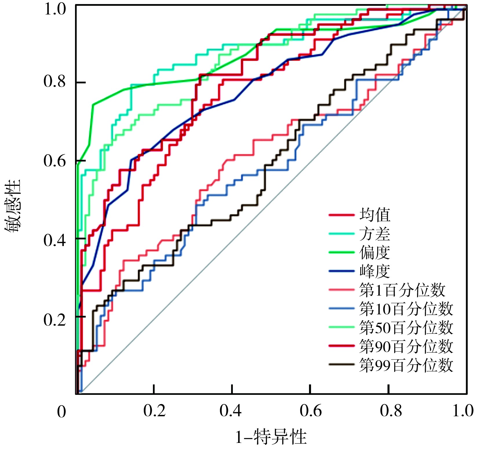
"
| 参数 | AUC(95%CI) | 敏感性 | 特异性 | 约登指数 | 最佳截断值 |
|---|---|---|---|---|---|
| 均值 | 0.77(0.68~0.85) | 0.81 | 0.66 | 0.47 | 109.72 |
| 方差 | 0.88(0.82~0.97) | 0.80 | 0.87 | 0.67 | 1 136.35 |
| 偏度 | 0.88(0.80~0.95) | 0.75 | 0.96 | 0.71 | 0.16 |
| 峰度 | 0.78(0.71~0.86) | 0.60 | 0.88 | 0.48 | 0.18 |
| 第1百分位数 | 0.61(0.55~0.68) | 0.35 | 0.89 | 0.24 | 34.06 |
| 第10百分位数 | 0.58(0.52~0.66) | 0.50 | 0.69 | 0.19 | 62.45 |
| 第50百分位数 | 0.86(0.81~0.94) | 0.65 | 0.94 | 0.59 | 104.21 |
| 第90百分位数 | 0.82(0.74~0.91) | 0.82 | 0.69 | 0.51 | 147.65 |
| 第99百分位数 | 0.60(0.54~0.68) | 0.22 | 0.95 | 0.17 | 177.41 |
"
| 因素 | β值 | SE值 | Wald χ2值 | OR值 | 95%CI | P值 |
|---|---|---|---|---|---|---|
| 年龄(>57.99岁/≤57.99岁) | 2.30 | 1.126 | 4.17 | 9.98 | 1.10~90.70 | 0.041 |
| 肿瘤最大径(>2 cm /≤2 cm) | 2.00 | 0.980 | 4.15 | 7.36 | 1.08~50.23 | 0.042 |
| 内镜分型(Ⅲ型/Ⅰ型+Ⅱ型) | 1.79 | 1.610 | 1.24 | 5.99 | 0.26~140.48 | 0.266 |
| 组织分型(黏液腺癌/腺癌+其他) | 2.01 | 2.681 | 0.56 | 7.46 | 0.39~142.94 | 0.453 |
| 分化程度(低分化/高、中分化) | 2.99 | 1.430 | 4.37 | 19.88 | 1.21~327.92 | 0.037 |
| 均值(>109.72/≤109.72) | 2.00 | 1.022 | 3.82 | 7.37 | 0.99~54.68 | 0.051 |
| 方差(>1 136.35/≤1 136.35) | 2.79 | 1.006 | 7.67 | 16.24 | 2.26~116.68 | 0.006 |
| 偏度(>0.16/≤0.16) | 3.05 | 1.032 | 8.74 | 21.13 | 2.80~59.61 | 0.003 |
| 峰度(>0.18/≤0.18) | 1.35 | 0.965 | 1.96 | 3.86 | 0.58~25.62 | 0.162 |
| 第1百分位数(>34.06/≤34.06) | 2.28 | 1.083 | 4.43 | 9.78 | 1.17~81.76 | 0.035 |
| 第10百分位数(>62.45/≤62.45) | 1.52 | 1.054 | 2.09 | 4.58 | 0.58~36.13 | 0.149 |
| 第50百分位数(>104.21/≤104.21) | 1.69 | 0.953 | 3.16 | 5.44 | 0.84~35.21 | 0.075 |
| 第90百分位数(>147.65/≤147.65) | 1.75 | 0.999 | 3.08 | 5.78 | 0.82~40.91 | 0.079 |
| 第99百分位数(>177.41/≤177.41) | -0.16 | 0.906 | 0.03 | 0.85 | 0.15~5.05 | 0.862 |
"
| [1] |
Jacobsson M, Wagner V, Kanneganti S. Screening for colorectal cancer[J]. Surg Clin North Am, 2024, 104(3): 595-607. DOI: 10.1016/j.suc.2023.11.009.
pmid: 38677823 |
| [2] | Pinto RA, Kawaguti FS, Kimura CMS, et al. Comparing three-dimensional endorectal ultrasound and magnification chromoendoscopy for early rectal neoplasia invasion depth assessment[J]. J Gastroenterol Hepatol, 2024, 39(2): 346-352. DOI: 10.1111/jgh.16382. |
| [3] | Zhu HB, Zhao B, Li XT, et al. Value of multiple models of diffusion-weighted imaging to predict hepatic lymph node metastases in colorectal liver metastases patients[J]. World J Gastroenterol, 2024, 30(4): 308-317. DOI: 10.3748/wjg.v30.i4.308. |
| [4] | Romeo V, Kapetas P, Clauser P, et al. Simultaneous 18F-FDG PET/MRI radiomics and machine learning analysis of the primary breast tumor for the preoperative prediction of axillary lymph node status in breast cancer[J]. Cancers (Basel), 2023, 15(20): 5088. DOI: 10.3390/cancers15205088. |
| [5] | Kowal P, Ratajczyk K, Bursiewicz W, et al. Differentiation of solid and friable tumour thrombus in patients with renal cell carcinoma: the role of MRI apparent diffusion coefficient[J]. Adv Med Sci, 2024, 69(2): 434-442. DOI: 10.1016/j.advms.2024.09.002. |
| [6] |
Anh DV, Lam TV, Anh NTH, et al. Assessment of histogram analysis in distinguishing between low-grade and high-grade brainstem glioma[J]. Clin Ter, 2024, 175(5): 296-306. DOI: 10.7417/CT.2024.5134.
pmid: 39400094 |
| [7] | 中华医学会消化内镜学分会消化系早癌内镜诊断与治疗协作组, 中华医学会消化病学分会消化道肿瘤协作组, 中华医学会消化内镜学分会肠道学组, 等. 中国早期结直肠癌及癌前病变筛查与诊治共识[J]. 中国医刊, 2015, 50(2): 14-30. DOI: 10.3969/j.issn.1008-1070.2015.02.007. |
| [8] | Matsuura N, Kato M, Iwata K, et al. Endoscopic ultrasound classification for prediction of endoscopic submucosal dissection resectability: PREDICT classification[J]. Endosc Int Open, 2024, 12(9): E1075-E1084. DOI: 10.1055/a-2387-1754. |
| [9] | 冯广龙, 姜慧杰. ROC曲线分析在医学影像学诊断中的价值[J]. 中华医学杂志, 2015, 95(3): 231-233. DOI: 10.3760/cma.j.issn.0376-2491.2015.03.018. |
| [10] | Sharma M, Gupta A, Jana M, et al. Comparison of HASTE versus EPI-based DWI for retinoblastoma and correlation with prognostic histopathologic parameters[J]. AJNR Am J Neuroradiol, 2024, 45(2): 198-204. DOI: 10.3174/ajnr.A8084. |
| [11] | Gupta S, May FP, Kupfer SS, et al. Birth cohort colorectal cancer (CRC): Implications for research and practice[J]. Clin Gastroenterol Hepatol, 2024, 22(3): 455-469.e7. DOI: 10.1016/j.cgh.2023.11.040. |
| [12] | 唐进亮, 张爱国, 张岚, 等. 窄带成像技术结合超声内镜检查在结直肠癌浸润深度诊断中的应用[J]. 中国医疗设备, 2023, 38(8): 74-78. DOI: 10.3969/j.issn.1674-1633.2023.08.014. |
| [13] |
Dadgarnia M, Meybodian M, Mandegari M, et al. The relationship between depth of invasion and cervical lymph node metastasis in patients with laryngeal squamous cell carcinoma[J]. Eur Arch Otorhinolaryngol, 2025, 282(3): 1375-1379. DOI: 10.1007/s00405-025-09215-0.
pmid: 39915314 |
| [14] | 魏重操, 王晶, 邢欣, 等. 早期结直肠癌的诊断率及临床病理特征分析[J]. 胃肠病学和肝病学杂志, 2022, 31(10): 1154-1159. DOI: 10.3969/j.issn.1006-5709.2022.10.016. |
| [15] |
Baykara M, Yildirim M. Differentiation of multiple myeloma and metastases with apparent diffusion coefficient map histogram analysis[J]. North Clin Istanb, 2022, 9(3): 256-260. DOI: 10.14744/nci.2021.59376.
pmid: 36199864 |
| [16] | Cao T, Jiang R, Zheng L, et al. T1 and ADC histogram parameters may be an in vivo biomarker for predicting the grade, subtype, and proliferative activity of meningioma[J]. Eur Radiol, 2023, 33(1): 258-269. DOI: 10.1007/s00330-022-09026-5. |
| [17] | Tsuchiya H, Matoba M, Nishino Y, et al. Clinical utility of combined assessments of 4D volumetric perfusion CT, diffusion-weighted MRI and 18F-FDG PET-CT for the prediction of outcomes of head and neck squamous cell carcinoma treated with chemoradiotherapy[J]. Radiat Oncol, 2023, 18(1): 24. DOI: 10.1186/s13014-023-02202-x. |
| [18] | Deng L, Yang J, Zhang M, et al. Whole-lesion iodine map histogram analysis versus single-slice spectral CT parameters for determining novel International Association for the Study of Lung Cancer grade of invasive non-mucinous pulmonary adenocarcinomas[J]. Diagn Interv Imaging, 2024, 105(5): 165-173. DOI: 10.1016/j.diii.2023.12.001. |
| [19] | Gihr G, Horvath-Rizea D, Kohlhof-Meinecke P, et al. Diffusion weighted imaging in gliomas: a histogram-based approach for tumor characterization[J]. Cancers (Basel), 2022, 14(14): 3393. DOI: 10.3390/cancers14143393. |
| [20] |
Wan L, Peng W, Zou S, et al. Predicting perineural invasion using histogram analysis of zoomed EPI diffusion-weighted imaging in rectal cancer[J]. Abdom Radiol (NY), 2022, 47(10): 3353-3363. DOI: 10.1007/s00261-022-03579-1.
pmid: 35779094 |
| Viewed | ||||||
|
Full text |
|
|||||
|
Abstract |
|
|||||