
Journal of International Oncology ›› 2025, Vol. 52 ›› Issue (10): 621-627.doi: 10.3760/cma.j.cn371439-20250123-00106
• Original Article • Previous Articles Next Articles
Ji Shengchao, Jin Xiaofeng, Ye Daixi, Lu Zehua, Xuan Lulu, Geng Chengjun( )
)
Received:2025-01-23
Revised:2025-07-31
Online:2025-10-08
Published:2025-11-12
Contact:
Geng Chengjun
E-mail:hfgcj@hotmail.com
Supported by:Ji Shengchao, Jin Xiaofeng, Ye Daixi, Lu Zehua, Xuan Lulu, Geng Chengjun. Diagnostic value of preoperative diffusion weighted imaging histogram parameters in the depth of invasion of early rectal cancer[J]. Journal of International Oncology, 2025, 52(10): 621-627.

"
"
"
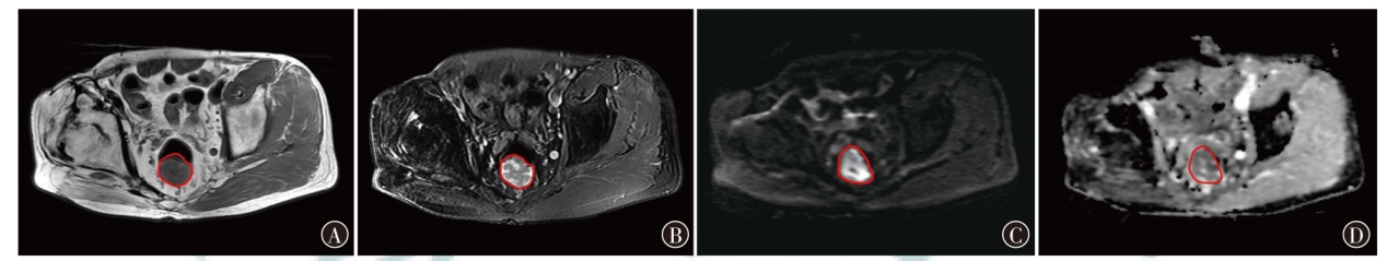
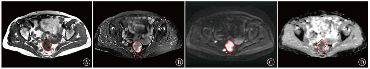
| 一般资料 | 黏膜内癌组 (n=102) | 黏膜下癌组 (n=78) | χ2/t值 | P值 |
|---|---|---|---|---|
| 性别 | ||||
| 男 | 49(48.04) | 44(56.41) | 1.24 | 0.265 |
| 女 | 53(51.96) | 34(43.59) | ||
| 年龄(岁) | 54.18±6.87 | 62.97±7.54 | 8.15 | <0.001 |
| BMI(kg/m2) | 23.87±3.98 | 24.02±3.52 | 0.26 | 0.793 |
| 吸烟史 | ||||
| 无 | 68(66.67) | 46(58.97) | 1.13 | 0.289 |
| 有 | 34(33.33) | 32(41.03) | ||
| 饮酒史 | ||||
| 无 | 56(54.90) | 38(48.72) | 0.68 | 0.410 |
| 有 | 46(45.10) | 40(51.28) | ||
| 家族史 | ||||
| 无 | 76(74.51) | 54(69.23) | 0.61 | 0.443 |
| 有 | 26(25.49) | 24(30.77) | ||
| 肿瘤最大径(cm) | ||||
| ≤2.0 | 74(72.55) | 25(32.05) | 29.29 | <0.001 |
| >2.0 | 28(27.45) | 53(67.95) | ||
| 内镜分型 | ||||
| Ⅰ型 | 72(70.59) | 34(43.59) | ||
| Ⅱ型 | 24(23.53) | 21(26.92) | 20.96 | <0.001 |
| Ⅲ型 | 6(5.88) | 23(29.49) | ||
| 组织分型 | ||||
| 腺癌 | 87(85.29) | 48(61.54) | ||
| 黏液腺癌 | 2(1.96) | 21(26.92) | 24.93 | <0.001 |
| 其他 | 13(12.75) | 9(11.54) | ||
| 分化程度 | ||||
| 高分化 | 78(76.47) | 13(16.67) | ||
| 中分化 | 20(19.61) | 24(30.77) | 75.35 | <0.001 |
| 低分化 | 4(3.92) | 41(52.56) |
"
| DWI直方图参数 | ICC | 95%CI |
|---|---|---|
| 均值 | 0.80 | 0.79~0.82 |
| 方差 | 0.92 | 0.91~0.94 |
| 偏度 | 0.91 | 0.89~0.92 |
| 峰度 | 0.91 | 0.90~0.93 |
| 第1百分位数 | 0.90 | 0.87~0.92 |
| 第10百分位数 | 0.88 | 0.86~0.91 |
| 第50百分位数 | 0.90 | 0.89~0.93 |
| 第90百分位数 | 0.85 | 0.82~0.97 |
| 第99百分位数 | 0.79 | 0.77~0.81 |
"
| 参数 | 黏膜内癌组 (n=102) | 黏膜下癌组 (n=78) | t值 | P值 |
|---|---|---|---|---|
| 均值 | 102.64±18.64 | 118.97±19.68 | 5.69 | <0.001 |
| 方差 | 1 022.34±162.31 | 1 285.44±199.64 | 9.75 | <0.001 |
| 偏度 | 0.12±0.04 | 0.21±0.07 | 10.88 | <0.001 |
| 峰度 | 0.14±0.05 | 0.23±0.07 | 10.06 | <0.001 |
| 第1百分位数 | 32.47±6.54 | 36.15±7.84 | 3.43 | <0.001 |
| 第10百分位数 | 60.35±8.67 | 65.19±9.32 | 3.59 | <0.001 |
| 第50百分位数 | 93.18±15.05 | 118.64±19.22 | 9.97 | <0.001 |
| 第90百分位数 | 141.28±20.31 | 155.97±22.08 | 4.63 | <0.001 |
| 第99百分位数 | 173.64±22.98 | 182.34±24.65 | 2.44 | 0.016 |
"
"
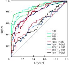
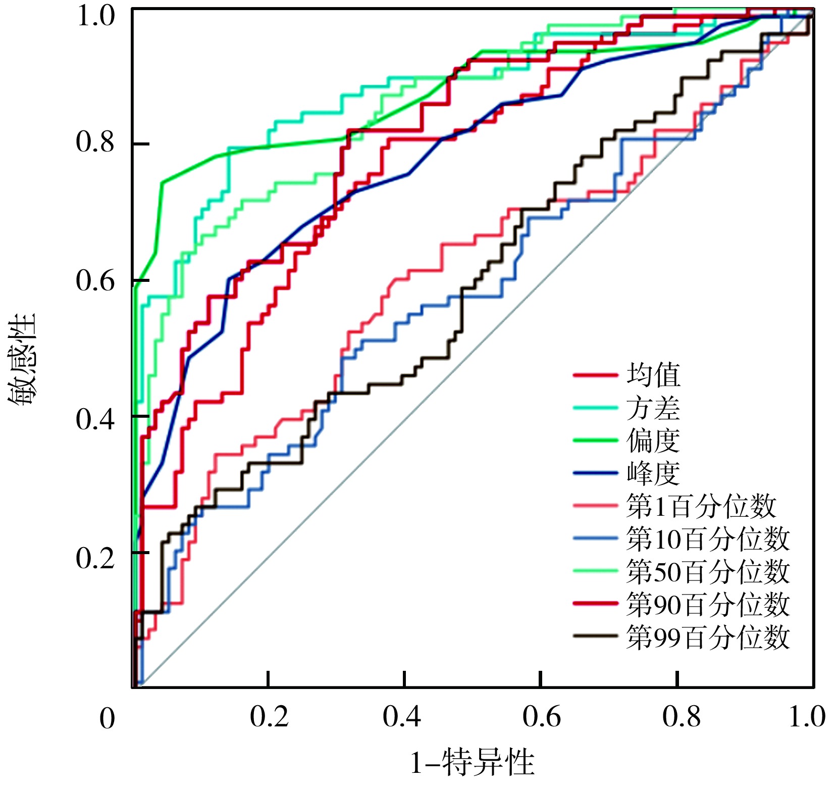
| 参数 | AUC(95%CI) | 敏感性 | 特异性 | 约登指数 | 最佳截断值 |
|---|---|---|---|---|---|
| 均值 | 0.77(0.68~0.85) | 0.81 | 0.66 | 0.47 | 109.72 |
| 方差 | 0.88(0.82~0.97) | 0.80 | 0.87 | 0.67 | 1 136.35 |
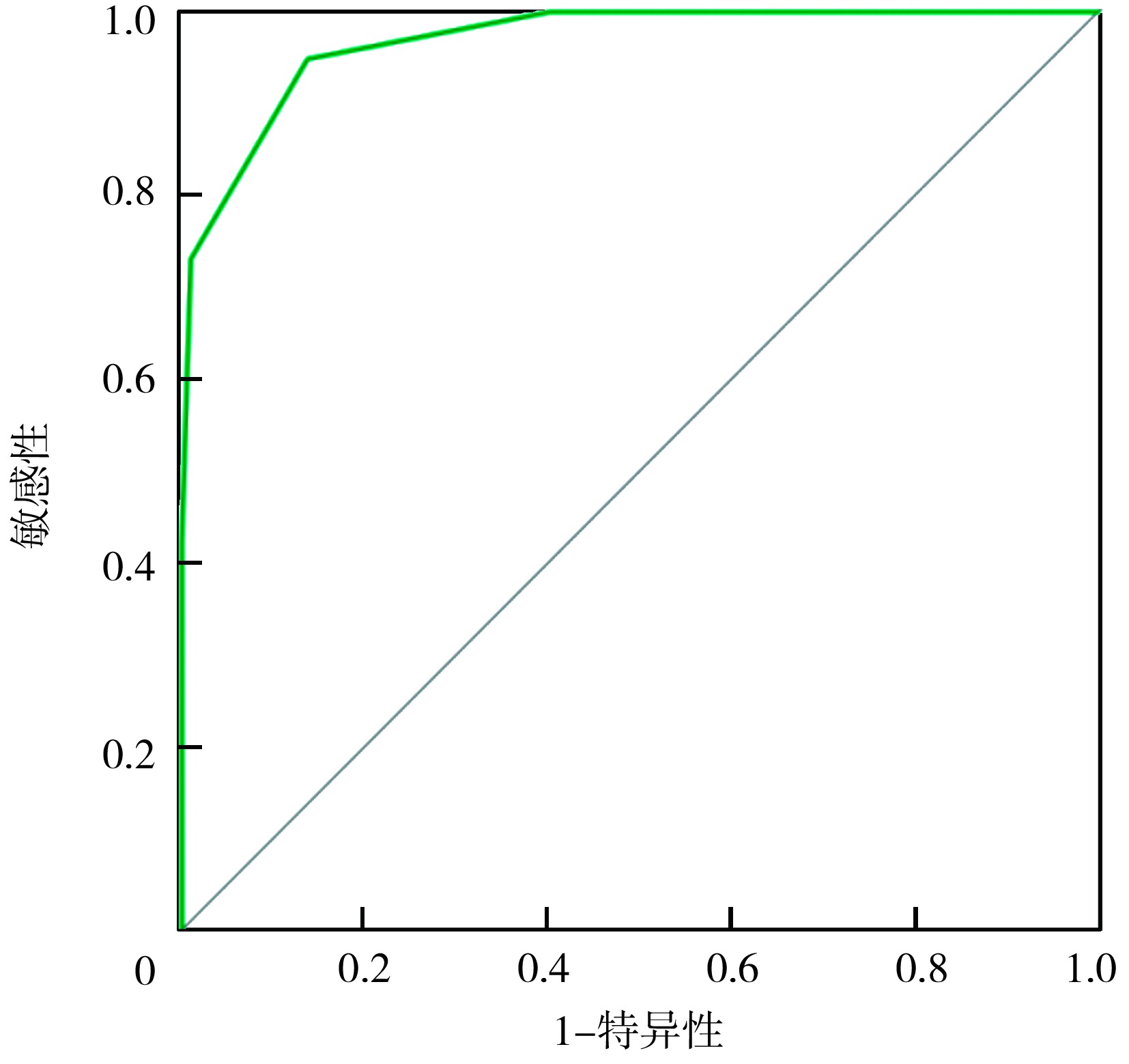
| 偏度 | 0.88(0.80~0.95) | 0.75 | 0.96 | 0.71 | 0.16 |
| 峰度 | 0.78(0.71~0.86) | 0.60 | 0.88 | 0.48 | 0.18 |
| 第1百分位数 | 0.61(0.55~0.68) | 0.35 | 0.89 | 0.24 | 34.06 |
| 第10百分位数 | 0.58(0.52~0.66) | 0.50 | 0.69 | 0.19 | 62.45 |
| 第50百分位数 | 0.86(0.81~0.94) | 0.65 | 0.94 | 0.59 | 104.21 |
| 第90百分位数 | 0.82(0.74~0.91) | 0.82 | 0.69 | 0.51 | 147.65 |
| 第99百分位数 | 0.60(0.54~0.68) | 0.22 | 0.95 | 0.17 | 177.41 |
"
| 因素 | β值 | SE值 | Wald χ2值 | OR值 | 95%CI | P值 |
|---|---|---|---|---|---|---|
| 年龄(>57.99岁/≤57.99岁) | 2.30 | 1.126 | 4.17 | 9.98 | 1.10~90.70 | 0.041 |
| 肿瘤最大径(>2 cm /≤2 cm) | 2.00 | 0.980 | 4.15 | 7.36 | 1.08~50.23 | 0.042 |
| 内镜分型(Ⅲ型/Ⅰ型+Ⅱ型) | 1.79 | 1.610 | 1.24 | 5.99 | 0.26~140.48 | 0.266 |
| 组织分型(黏液腺癌/腺癌+其他) | 2.01 | 2.681 | 0.56 | 7.46 | 0.39~142.94 | 0.453 |
| 分化程度(低分化/高、中分化) | 2.99 | 1.430 | 4.37 | 19.88 | 1.21~327.92 | 0.037 |
| 均值(>109.72/≤109.72) | 2.00 | 1.022 | 3.82 | 7.37 | 0.99~54.68 | 0.051 |
| 方差(>1 136.35/≤1 136.35) | 2.79 | 1.006 | 7.67 | 16.24 | 2.26~116.68 | 0.006 |
| 偏度(>0.16/≤0.16) | 3.05 | 1.032 | 8.74 | 21.13 | 2.80~59.61 | 0.003 |
| 峰度(>0.18/≤0.18) | 1.35 | 0.965 | 1.96 | 3.86 | 0.58~25.62 | 0.162 |
| 第1百分位数(>34.06/≤34.06) | 2.28 | 1.083 | 4.43 | 9.78 | 1.17~81.76 | 0.035 |
| 第10百分位数(>62.45/≤62.45) | 1.52 | 1.054 | 2.09 | 4.58 | 0.58~36.13 | 0.149 |
| 第50百分位数(>104.21/≤104.21) | 1.69 | 0.953 | 3.16 | 5.44 | 0.84~35.21 | 0.075 |
| 第90百分位数(>147.65/≤147.65) | 1.75 | 0.999 | 3.08 | 5.78 | 0.82~40.91 | 0.079 |
| 第99百分位数(>177.41/≤177.41) | -0.16 | 0.906 | 0.03 | 0.85 | 0.15~5.05 | 0.862 |
"
| [1] |
Jacobsson M, Wagner V, Kanneganti S. Screening for colorectal cancer[J]. Surg Clin North Am, 2024, 104(3): 595-607. DOI: 10.1016/j.suc.2023.11.009.
pmid: 38677823 |
| [2] | Pinto RA, Kawaguti FS, Kimura CMS, et al. Comparing three-dimensional endorectal ultrasound and magnification chromoendoscopy for early rectal neoplasia invasion depth assessment[J]. J Gastroenterol Hepatol, 2024, 39(2): 346-352. DOI: 10.1111/jgh.16382. |
| [3] | Zhu HB, Zhao B, Li XT, et al. Value of multiple models of diffusion-weighted imaging to predict hepatic lymph node metastases in colorectal liver metastases patients[J]. World J Gastroenterol, 2024, 30(4): 308-317. DOI: 10.3748/wjg.v30.i4.308. |
| [4] | Romeo V, Kapetas P, Clauser P, et al. Simultaneous 18F-FDG PET/MRI radiomics and machine learning analysis of the primary breast tumor for the preoperative prediction of axillary lymph node status in breast cancer[J]. Cancers (Basel), 2023, 15(20): 5088. DOI: 10.3390/cancers15205088. |
| [5] | Kowal P, Ratajczyk K, Bursiewicz W, et al. Differentiation of solid and friable tumour thrombus in patients with renal cell carcinoma: the role of MRI apparent diffusion coefficient[J]. Adv Med Sci, 2024, 69(2): 434-442. DOI: 10.1016/j.advms.2024.09.002. |
| [6] |
Anh DV, Lam TV, Anh NTH, et al. Assessment of histogram analysis in distinguishing between low-grade and high-grade brainstem glioma[J]. Clin Ter, 2024, 175(5): 296-306. DOI: 10.7417/CT.2024.5134.
pmid: 39400094 |
| [7] | 中华医学会消化内镜学分会消化系早癌内镜诊断与治疗协作组, 中华医学会消化病学分会消化道肿瘤协作组, 中华医学会消化内镜学分会肠道学组, 等. 中国早期结直肠癌及癌前病变筛查与诊治共识[J]. 中国医刊, 2015, 50(2): 14-30. DOI: 10.3969/j.issn.1008-1070.2015.02.007. |
| [8] | Matsuura N, Kato M, Iwata K, et al. Endoscopic ultrasound classification for prediction of endoscopic submucosal dissection resectability: PREDICT classification[J]. Endosc Int Open, 2024, 12(9): E1075-E1084. DOI: 10.1055/a-2387-1754. |
| [9] | 冯广龙, 姜慧杰. ROC曲线分析在医学影像学诊断中的价值[J]. 中华医学杂志, 2015, 95(3): 231-233. DOI: 10.3760/cma.j.issn.0376-2491.2015.03.018. |
| [10] | Sharma M, Gupta A, Jana M, et al. Comparison of HASTE versus EPI-based DWI for retinoblastoma and correlation with prognostic histopathologic parameters[J]. AJNR Am J Neuroradiol, 2024, 45(2): 198-204. DOI: 10.3174/ajnr.A8084. |
| [11] | Gupta S, May FP, Kupfer SS, et al. Birth cohort colorectal cancer (CRC): Implications for research and practice[J]. Clin Gastroenterol Hepatol, 2024, 22(3): 455-469.e7. DOI: 10.1016/j.cgh.2023.11.040. |
| [12] | 唐进亮, 张爱国, 张岚, 等. 窄带成像技术结合超声内镜检查在结直肠癌浸润深度诊断中的应用[J]. 中国医疗设备, 2023, 38(8): 74-78. DOI: 10.3969/j.issn.1674-1633.2023.08.014. |
| [13] |
Dadgarnia M, Meybodian M, Mandegari M, et al. The relationship between depth of invasion and cervical lymph node metastasis in patients with laryngeal squamous cell carcinoma[J]. Eur Arch Otorhinolaryngol, 2025, 282(3): 1375-1379. DOI: 10.1007/s00405-025-09215-0.
pmid: 39915314 |
| [14] | 魏重操, 王晶, 邢欣, 等. 早期结直肠癌的诊断率及临床病理特征分析[J]. 胃肠病学和肝病学杂志, 2022, 31(10): 1154-1159. DOI: 10.3969/j.issn.1006-5709.2022.10.016. |
| [15] |
Baykara M, Yildirim M. Differentiation of multiple myeloma and metastases with apparent diffusion coefficient map histogram analysis[J]. North Clin Istanb, 2022, 9(3): 256-260. DOI: 10.14744/nci.2021.59376.
pmid: 36199864 |
| [16] | Cao T, Jiang R, Zheng L, et al. T1 and ADC histogram parameters may be an in vivo biomarker for predicting the grade, subtype, and proliferative activity of meningioma[J]. Eur Radiol, 2023, 33(1): 258-269. DOI: 10.1007/s00330-022-09026-5. |
| [17] | Tsuchiya H, Matoba M, Nishino Y, et al. Clinical utility of combined assessments of 4D volumetric perfusion CT, diffusion-weighted MRI and 18F-FDG PET-CT for the prediction of outcomes of head and neck squamous cell carcinoma treated with chemoradiotherapy[J]. Radiat Oncol, 2023, 18(1): 24. DOI: 10.1186/s13014-023-02202-x. |
| [18] | Deng L, Yang J, Zhang M, et al. Whole-lesion iodine map histogram analysis versus single-slice spectral CT parameters for determining novel International Association for the Study of Lung Cancer grade of invasive non-mucinous pulmonary adenocarcinomas[J]. Diagn Interv Imaging, 2024, 105(5): 165-173. DOI: 10.1016/j.diii.2023.12.001. |
| [19] | Gihr G, Horvath-Rizea D, Kohlhof-Meinecke P, et al. Diffusion weighted imaging in gliomas: a histogram-based approach for tumor characterization[J]. Cancers (Basel), 2022, 14(14): 3393. DOI: 10.3390/cancers14143393. |
| [20] |
Wan L, Peng W, Zou S, et al. Predicting perineural invasion using histogram analysis of zoomed EPI diffusion-weighted imaging in rectal cancer[J]. Abdom Radiol (NY), 2022, 47(10): 3353-3363. DOI: 10.1007/s00261-022-03579-1.
pmid: 35779094 |
| Viewed | ||||||
|
Full text |
|
|||||
|
Abstract |
|
|||||