Journal of International Oncology ›› 2025, Vol. 52 ›› Issue (3): 136-143.doi: 10.3760/cma.j.cn371439-20241021-00021
• Original Article • Previous Articles Next Articles
CT feature analysis and predictive value of visceral pleural invasion in stage Ⅰ lung adenocarcinoma with peripheral solid nodules
Han Shuang( )
)
- Department of Radiology,Second Affiliated Hospital of Guizhou University of Traditional Chinese Medicine,Guiyang 550003,China
-
Received:2024-10-21Revised:2024-12-19Online:2025-03-08Published:2025-04-02 -
Contact:Email:1527698862@qq.com

Cite this article
Han Shuang. CT feature analysis and predictive value of visceral pleural invasion in stage Ⅰ lung adenocarcinoma with peripheral solid nodules[J]. Journal of International Oncology, 2025, 52(3): 136-143.
share this article

"
| 项目 | 训练集(n=112) | 验证集(n=38) | t/χ2值 | P值 | 项目 | 训练集(n=112) | 验证集(n=38) | t/χ2值 | P值 |
|---|---|---|---|---|---|---|---|---|---|
| 年龄(岁) | 60.30±10.19 | 59.52±9.69 | 0.41 | 0.680 | 钙化 | ||||
| 性别 | 有 | 6 | 5 | 1.52 | 0.217 | ||||
| 男 | 44 | 20 | 2.07 | 0.151 | 无 | 106 | 33 | ||
| 女 | 68 | 18 | 瘤内坏死 | ||||||
| 病理类型 | 有 | 31 | 8 | 0.65 | 0.421 | ||||
| AIS | 20 | 6 | 0.16 | 0.921 | 无 | 81 | 30 | ||
| MIA | 18 | 7 | 空泡征 | ||||||
| IA | 74 | 25 | 有 | 27 | 4 | 3.19 | 0.074 | ||
| 病灶分布 | 无 | 85 | 34 | ||||||
| 右肺 | 70 | 24 | 0.01 | 0.942 | 空气支气管征 | ||||
| 左肺上叶 | 42 | 14 | 有 | 31 | 12 | 0.21 | 0.646 | ||
| VPI | 无 | 81 | 26 | ||||||
| 阳性 | 35 | 12 | <0.01 | 0.970 | 周围性肺气肿 | ||||
| 阴性 | 77 | 26 | 有 | 11 | 7 | 1.26 | 0.262 | ||
| 病灶最大径(cm) | 1.71±0.43 | 1.75±0.35 | 0.52 | 0.605 | 无 | 101 | 31 | ||
| 形态 | 外周纤维化 | ||||||||
| 规则 | 39 | 13 | 0.01 | 0.945 | 有 | 27 | 8 | 0.15 | 0.700 |
| 不规则 | 73 | 25 | 无 | 85 | 30 | ||||
| 边界 | 胸膜牵拉 | ||||||||
| 清晰 | 72 | 24 | 0.02 | 0.900 | 有 | 65 | 25 | 0.71 | 0.399 |
| 模糊 | 40 | 14 | 无 | 47 | 13 | ||||
| 分叶征 | 胸膜接触 | ||||||||
| 有 | 81 | 23 | 1.86 | 0.173 | 有 | 41 | 10 | 1.34 | 0.247 |
| 无 | 31 | 15 | 无 | 71 | 28 | ||||
| 毛刺征 | 邻近胸膜增厚 | ||||||||
| 有 | 63 | 22 | 0.03 | 0.860 | 有 | 38 | 13 | <0.01 | 0.975 |
| 无 | 49 | 16 | 无 | 74 | 25 | ||||
| 密度 | 淋巴结肿大 | ||||||||
| 实性 | 48 | 19 | 0.59 | 0.444 | 有 | 13 | 3 | 1.10 | 0.736 |
| 亚实性 | 64 | 19 | 无 | 99 | 35 | ||||
| 晕征 | |||||||||
| 有 | 53 | 20 | 0.32 | 0.571 | |||||
| 无 | 59 | 18 |
"
| 项目 | VPI阴性组(n=77) | VPI阳性组(n=35) | t/χ2值 | P值 | 项目 | VPI阴性组(n=77) | VPI阳性组(n=35) | t/χ2值 | P值 |
|---|---|---|---|---|---|---|---|---|---|
| 年龄(岁) | 59.27±11.18 | 61.15±8.82 | 0.88 | 0.382 | 钙化 | ||||
| 性别 | 有 | 3 | 3 | 0.32 | 0.571 | ||||
| 男 | 27 | 17 | 1.84 | 0.175 | 无 | 74 | 32 | ||
| 女 | 50 | 18 | 瘤内坏死 | ||||||
| 病理类型 | 有 | 16 | 15 | 5.86 | 0.015 | ||||
| AIS | 20 | 0 | 11.49 | 0.003 | 无 | 61 | 20 | ||
| MIA | 10 | 8 | 空泡征 | ||||||
| IA | 47 | 27 | 有 | 17 | 10 | 0.56 | 0.456 | ||
| 病灶分布 | 无 | 60 | 25 | ||||||
| 右肺 | 49 | 21 | 0.14 | 0.713 | 空气支气管征 | ||||
| 左肺 | 28 | 14 | 有 | 18 | 13 | 2.28 | 0.131 | ||
| 病灶最大径(cm) | 1.50±0.44 | 2.12±0.67 | 5.83 | <0.001 | 无 | 59 | 22 | ||
| 形态 | 周围性肺气肿 | ||||||||
| 规则 | 29 | 10 | 0.88 | 0.349 | 有 | 5 | 6 | 3.08 | 0.079 |
| 不规则 | 48 | 25 | 无 | 72 | 29 | ||||
| 边界 | 外周纤维化 | ||||||||
| 清晰 | 50 | 22 | 0.05 | 0.832 | 有 | 16 | 11 | 1.49 | 0.222 |
| 模糊 | 27 | 13 | 无 | 61 | 24 | ||||
| 分叶征 | 胸膜牵拉 | ||||||||
| 有 | 49 | 32 | 9.29 | 0.002 | 有 | 36 | 29 | 12.88 | <0.001 |
| 无 | 28 | 3 | 无 | 41 | 6 | ||||
| 毛刺征 | 胸膜接触 | ||||||||
| 有 | 39 | 24 | 3.14 | 0.076 | 有 | 23 | 18 | 4.82 | 0.028 |
| 无 | 38 | 11 | 无 | 54 | 17 | ||||
| 密度 | 邻近胸膜增厚 | ||||||||
| 实性 | 26 | 22 | 8.32 | 0.004 | 有 | 21 | 17 | 4.87 | 0.027 |
| 亚实性 | 51 | 13 | 无 | 56 | 18 | ||||
| 晕征 | 淋巴结肿大 | ||||||||
| 有 | 33 | 20 | 1.97 | 0.160 | 有 | 8 | 5 | 0.08 | 0.781 |
| 无 | 44 | 15 | 无 | 69 | 30 |
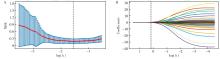
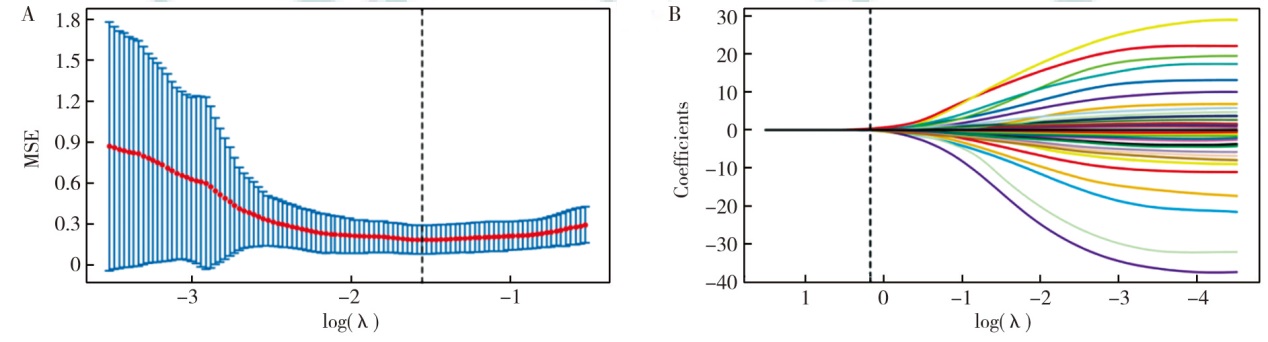
"
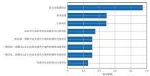
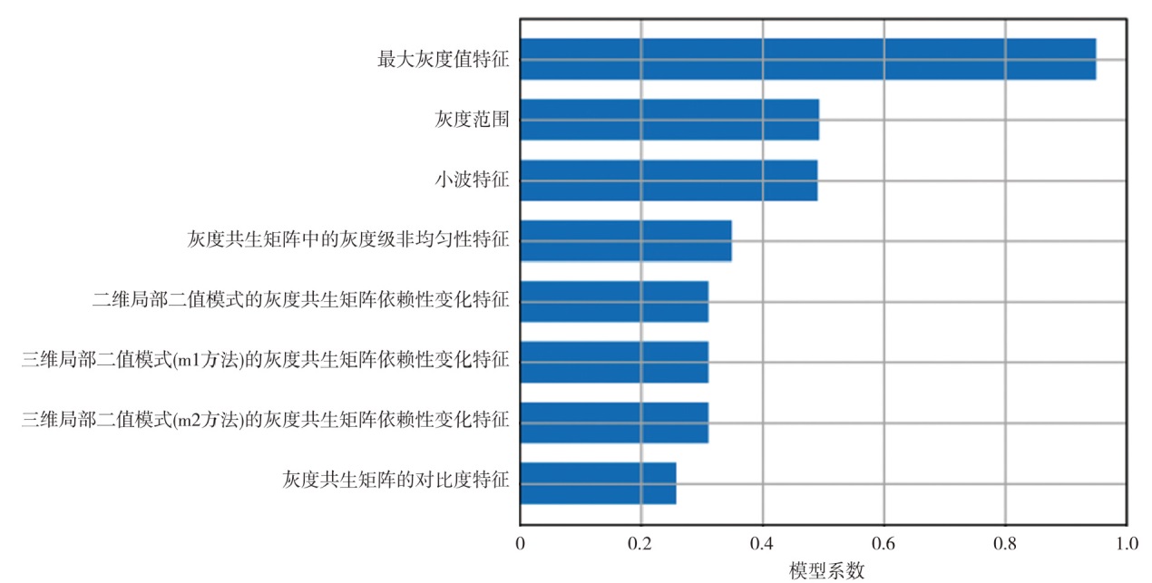
"
"
| 项目 | 单因素 | 多因素 | |||||
|---|---|---|---|---|---|---|---|
| OR值 | 95%CI | P值 | OR值 | 95%CI | P值 | ||
| 病理类型 | |||||||
| IA | Ref | ||||||
| AIS | 1.21 | 0.44~3.31 | 0.350 | ||||
| MIA | 1.02 | 0.97~1.05 | 0.685 | ||||
| 病灶最大径 | 1.48 | 1.09~2.01 | 0.010 | 1.94 | 0.33~3.18 | 0.151 | |
| 分叶征 | |||||||
| 无 | Ref | ||||||
| 有 | 5.09 | 2.31~6.00 | 0.001 | 6.42 | 1.42~18.58 | 0.018 | |
| 密度 | |||||||
| 亚实性 | Ref | ||||||
| 实性 | 4.25 | 1.47~7.18 | 0.004 | 0.74 | 0.23~2.33 | 0.930 | |
| 瘤内坏死 | |||||||
| 无 | Ref | ||||||
| 有 | 2.27 | 1.01~5.17 | 0.049 | 3.63 | 1.01~10.01 | 0.046 | |
| 胸膜牵拉 | |||||||
| 无 | Ref | ||||||
| 有 | 6.75 | 1.92~13.68 | <0.001 | 4.19 | 1.17~10.92 | 0.028 | |
| 胸膜接触 | |||||||
| 无 | Ref | ||||||
| 有 | 3.58 | 1.18~5.65 | 0.018 | 1.02 | 0.32~3.22 | 0.607 | |
| 邻近胸膜增厚 | |||||||
| 无 | Ref | ||||||
| 有 | 3.60 | 1.18~5.72 | 0.018 | 0.73 | 0.48~1.12 | 0.954 | |
| 影像组学评分 | 19 418.06 | 394.18~957 161.04 | <0.001 | 179 711.20 | 525.13~61 552 573.59 | <0.001 | |
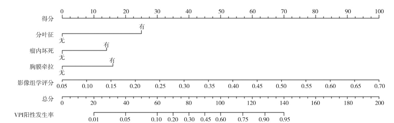
"
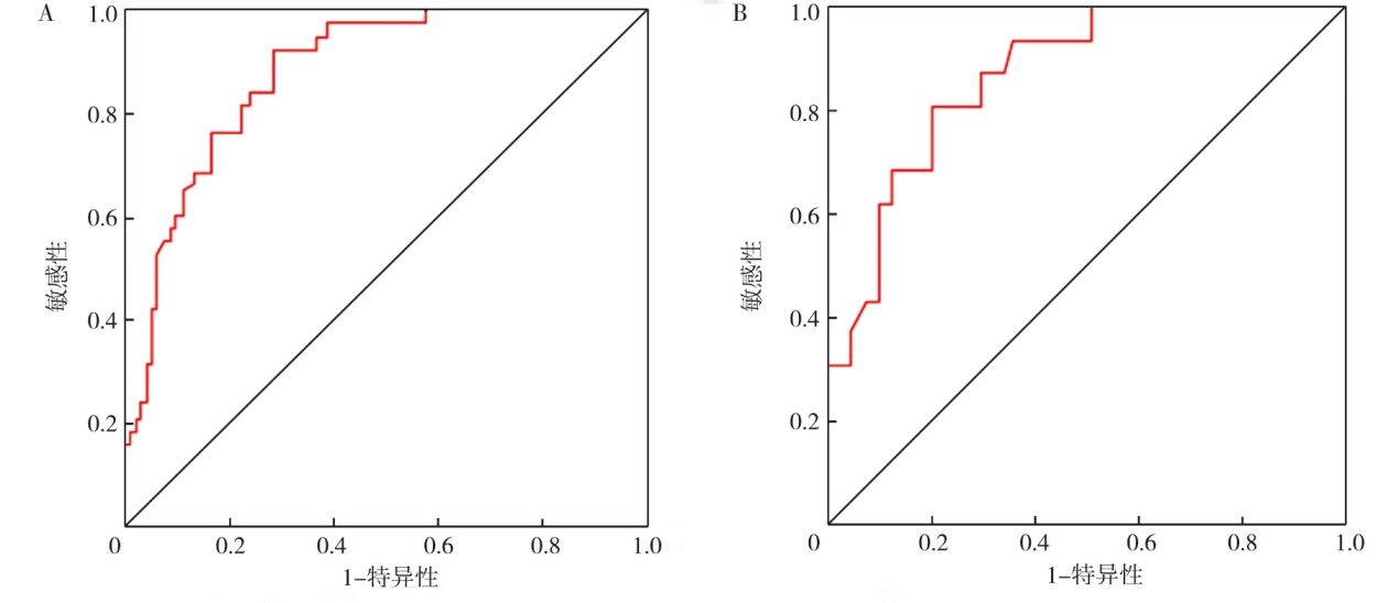
"
"
| [1] | Lim WH, Lee KH, Lee JH, et al. Diagnostic performance and prognostic value of CT-defined visceral pleural invasion in early-stage lung adenocarcinomas[J]. Eur Radiol, 2024, 34(3): 1934-1945. DOI: 10.1007/s00330-023-10204-2. |
| [2] | Nishino M. Jellyfish sign for visceral pleural invasion in lung cancer: reviving the lost art of radiology[J]. Radiology, 2024, 310(1): e233171. DOI: 10.1148/radiol.233171. |
| [3] | Yang L, Jiang Z, Tong J, et al. Development and validation of a preoperative CT-based radiomics nomogram to differentiate tuberculosis granulomas from lung adenocarcinomas: an external validation study[J]. BMC Cancer, 2024, 24(1): 670. DOI: 10.1186/s12885-024-12422-3. |
| [4] | Zhou H, Zhou J, Qin C, et al. Preoperative prediction of perineural invasion in oesophageal squamous cell carcinoma based on CT radiomics nomogram: a multicenter study[J]. Acad Radiol, 2024, 31(4): 1355-1366. DOI: 10.1016/j.acra.2023.09.026. |
| [5] |
Jin H, Wang Y, Li X, et al. Radiomics nomogram for predicting chemo-immunotherapy efficiency in advanced non-small cell lung cancer[J]. Sci Rep, 2024, 14(1): 20788. DOI: 10.1038/s41598-024-63415-y.
pmid: 39242619 |
| [6] | Li R, Peng H, Xue T, et al. Prediction and verification of survival in patients with non-small-cell lung cancer based on an integrated radiomics nomogram[J]. Clin Radiol, 2022, 77(3): e222-e230. DOI: 10.1016/j.crad.2021.12.002. |
| [7] |
Wang Y, Liu W, Yu Y, et al. CT radiomics nomogram for the preope-rative prediction of lymph node metastasis in gastric cancer[J]. Eur Radiol, 2020, 30(2): 976-986. DOI: 10.1007/s00330-019-06398-z.
pmid: 31468157 |
| [8] | Rami-Porta R, Asamura H, Travis WD, et al. Lung cancer-major changes in the American Joint Committee on Cancer eighth edition cancer staging manual[J]. CA Cancer J Clin, 2017, 67(2): 138-155. DOI: 10.3322/caac.21390. |
| [9] | 望云, 吕登, 涂文婷, 等. CT特征预测胸膜下临床ⅠA期周围型肺腺癌脏层胸膜侵犯的价值[J]. 中华放射学杂志, 2022, 56(10): 1103-1109. DOI: 10.3760/cma.j.cn112149-20220624-00540. |
| [10] | 朱浩楠, 王安生, 洪海宁, 等. 基于增强CT影像组学及形态学征象对非小细胞肺癌脏层胸膜侵犯的预测价值研究[J]. 中华解剖与临床杂志, 2022, 27(4): 213-219. DOI: 10.3760/cma.j.cn101202-20210803-00190. |
| [11] | Fang W, Zhang G, Yu Y, et al. Identification of pathological subtypes of early lung adenocarcinoma based on artificial intelligence para-meters and CT signs[J]. Biosci Rep, 2022, 42(1): BSR20212416. DOI: 10.1042/BSR20212416. |
| [12] | 苗嫚丽, 范晓云, 丁佩山, 等. 孤立性肺结节中Ⅰ期非小细胞肺癌的危险因素分析及其病理不同进展程度的临床特征对比[J]. 中国临床保健杂志, 2019, 22(6): 753-759. DOI: 10.3969/J.issn.1672-6790.2019.06.009. |
| [13] | 李鲁, 舒锦尔, 陈真伟, 等. 磨玻璃结节表现的肺腺癌侵犯脏层胸膜的超高分辨率CT评价[J]. 中国医学计算机成像杂志, 2020, 26(3): 223-229. DOI: 10.3969/j.issn.1006-5741.2020.03.006. |
| [14] | Onoda H, Higashi M, Murakami T, et al. Correlation between pleural tags on CT and visceral pleural invasion of peripheral lung cancer that does not appear touching the pleural surface[J]. Eur Radiol, 2021, 31(12): 9022-9029. DOI: 10.1007/s00330-021-07869-y. |
| [1] | Lai Ruihe, Teng Yue, Rong Jian, Sheng Dandan, Geng Yuzhi, Chen Jianxin, Jiang Chong, Ding Chongyang, Zhou Zhengyang. Predictive value of a combined model for lymph node metastasis in NSCLC based on primary lesion radiomics from 18F-FDG PET/CT [J]. Journal of International Oncology, 2025, 52(3): 144-151. |
| [2] | Yuan Shengfang, Ren Jie, Lin Weijia, Ji Zexuan, Zhang Changhong, Wang Bu. Prognostic value of EGFR co-mutation status in patients with advanced lung adenocarcinoma [J]. Journal of International Oncology, 2024, 51(9): 556-562. |
| [3] | Peng Dan, Lyu Lu, Sun Pengfei. Research progress of radiomics in cervical cancer [J]. Journal of International Oncology, 2024, 51(8): 532-537. |
| [4] | Fu Yi, Ma Chenying, Zhang Lu, Zhou Juying. Research progress of habitat analysis in radiomics of malignant tumors [J]. Journal of International Oncology, 2024, 51(5): 292-297. |
| [5] | Li Tian, Wu Yang, Zhang Jiangming, Xi Chunsheng. Construction of a nomogram prediction model for lung metastasis in elderly patients with clear cell renal cell carcinoma [J]. Journal of International Oncology, 2024, 51(12): 755-762. |
| [6] | Wang Xiao, Li Ying, Luo Yujie, Jin Shu. Study on the prognostic value of serological indicators for nasopharyngeal carcinoma based on nomogram model [J]. Journal of International Oncology, 2023, 50(8): 463-469. |
| [7] | Cao Xiaohui, Yu Hong, Li Wanhu. Application of CT-based radiomics analysis in predicting and identifying of treatment-associated pneumonitis [J]. Journal of International Oncology, 2023, 50(2): 107-111. |
| [8] | Liao Lihua, Wang Shaohong, Chen Hongcai, Zhao Yongqiang. Value of cell paraffin block immunohistochemistry and pleural effusion CRKL and MIC-1 in the diagnosis of malignant pleural effusion [J]. Journal of International Oncology, 2023, 50(2): 71-75. |
| [9] | Xie Yu, Zheng Shengnan, Huang Mingmin, Guo Aibin, Yin Zhenyu, Lin Yongjuan. Pemetrexed clinical trial for intrathecal injection chemotherapy based on cerebrospinal fluid pharmacokinetics in patients with leptomeningeal metastasis from lung adenocarcinoma [J]. Journal of International Oncology, 2023, 50(10): 585-591. |
| [10] | Wu Jiayu, Liu Jiacheng. Research progress of radiomics toward lung adenocarcinoma manifesting as solitary ground glass nodule [J]. Journal of International Oncology, 2022, 49(8): 449-452. |
| [11] | Li Yangyi, Gong Yan, Xie Conghua. SAMHD1 inhibits PD-L1 expression in lung adenocarcinoma cells [J]. Journal of International Oncology, 2022, 49(11): 658-664. |
| [12] | Yu Mingyue, Chen Zhengzheng, Zhao Xuxu, Ren Pingping, Zhang Ying, Ge Li, Zhu Meiling, Zhao Weidong. Factors related to postoperative adjuvant therapy of locally advanced cervical cancer and building of a nomogram prediction model [J]. Journal of International Oncology, 2021, 48(1): 35-40. |
| [13] | Yang Lei, Zhang Chuanyu, Zhang Zaixian, Liu Huan. Radiogenomics in non-small cell lung cancer [J]. Journal of International Oncology, 2020, 47(9): 555-559. |
| [14] | Guo Xinwei, Ji Shengjun, Ji Lei, Zhang Han, Zhou Shaobing, Liu Yangchen. Value of nomogram incorporated preoperative tumor volume on predicting the prognosis of thoracic esophageal squamous cell carcinoma patients [J]. Journal of International Oncology, 2020, 47(5): 278-283. |
| [15] | Lu Haidi, Shen Fu, Lu Jianping, Hao Liqiang. Research on prediction of pathological complete response after neoadjuvant therapy for rectal cancer based on MRI high-resolution T2WI images [J]. Journal of International Oncology, 2020, 47(10): 593-597. |
| Viewed | ||||||
|
Full text |
|
|||||
|
Abstract |
|
|||||