Journal of International Oncology ›› 2025, Vol. 52 ›› Issue (5): 282-287.doi: 10.3760/cma.j.cn371439-20240618-00048
• Original Article • Previous Articles Next Articles
Role of chemokine CX3CL1/CX3CR1 in intraperitoneal metastasis of ovarian cancer in nude mice
Zeng Qianqian, Xiang Hong( ), Fu Lijun
- Department of Obstetrics and Gynecology, First Affiliated Hospital of Xinjiang Medical University, Urumqi 830054, China
-
Received:2024-06-18Revised:2024-09-20Online:2025-05-08Published:2025-06-24 -
Contact:Xiang Hong E-mail:xianghong1965@163.com -
Supported by:Natural Science Foundation of Xinjiang Uygur Autonomous Region of China(81660288)
Cite this article
Zeng Qianqian, Xiang Hong, Fu Lijun. Role of chemokine CX3CL1/CX3CR1 in intraperitoneal metastasis of ovarian cancer in nude mice[J]. Journal of International Oncology, 2025, 52(5): 282-287.
share this article
"
| 组别 | 生存时间(d) |
|---|---|
| 正常组(n=10) | 14.00±0.00 |
| 卵巢癌模型组(n=20) | 9.24±0.67a |
| CX3CL1组(n=20) | 12.05±0.82ab |
| F值 | 22.27 |
| P值 | <0.001 |
"
| 组别 | 肿瘤质量(g) | 肿瘤体积(mm3) | 抑瘤率(%) | 腹腔积液 | 腹腔转移 |
|---|---|---|---|---|---|
| 卵巢癌模型组(n=20) | 1.31±0.21 | 130.47±13.45 | 0.00±0.00 | 12(60.00) | 16(80.00) |
| CX3CL1组(n=20) | 0.62±0.13 | 70.02±7.52 | 48.96±4.74 | 5(25.00) | 10(50.00) |
| t/χ2值 | 12.49 | 17.54 | 46.19 | 5.01 | 3.96 |
| P值 | <0.001 | <0.001 | <0.001 | 0.025 | 0.047 |
"

"
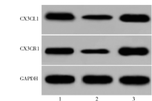
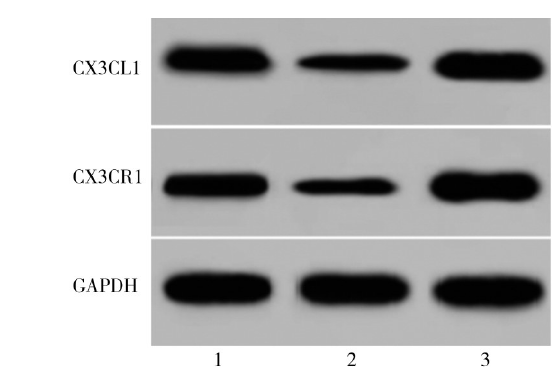
| 组别 | CX3CL1 | CX3CR1 |
|---|---|---|
| 正常组(n=10) | 2.05±0.22 | 1.99±0.21 |
| 卵巢癌模型组(n=20) | 1.33±0.11a | 1.34±0.14a |
| CX3CL1组(n=20) | 2.41±0.24ab | 2.73±0.31ab |
| F值 | 9.26 | 8.14 |
| P值 | <0.001 | <0.001 |
"
| [1] | Lumish MA, Kohn EC, Tew WP. Top advances of the year: ovarian cancer[J]. Cancer, 2024, 130(6): 837-845. DOI: 10.1002/cncr.35135. |
| [2] | 杨丽蓉, 王羽丰. 预测浆液性卵巢癌术后复发远处转移风险机器学习模型的构建[J]. 国际肿瘤学杂志, 2023, 50(4): 220-226. DOI: 10.3760/cma.j.cn371439-20221214-00043. |
| [3] | Nwabufo CK. Mirvetuximab soravtansine in ovarian cancer therapy: expert opinion on pharmacological considerations[J]. Cancer Chemother Pharmacol, 2024, 93(2): 89-105. DOI: 10.1007/s00280-023-04575-y. |
| [4] | Choi SY, Choi JH. Ovarian cancer and the microbiome: connecting the dots for early diagnosis and therapeutic innovations—a review[J]. Medicina (Kaunas), 2024, 60(3): 516. DOI: 10.3390/medicina 60030516. |
| [5] | Wong T, Tedja R, Chehade H, et al. An ex vivo model of ovarian cancer peritoneal metastasis using human omentum[J]. J Vis Exp, 2024, 26(203). DOI: 10.3791/66031. |
| [6] | Shao D, Zhou H, Yu H, et al. CX3CR1 is a potential biomarker of immune microenvironment and prognosis in epithelial ovarian cancer[J]. Medicine (Baltimore), 2024, 103(3): e36891. DOI: 10.1097/MD.0000000000036891. |
| [7] | Wang XC, Zhou H, Jiang WJ, et al. Effect of CX3CL1/CX3CR1 gene polymorphisms on the clinical efficacy of carboplatin therapy in Han patients with ovarian cancer[J]. Front Genet, 2022, 13: 1065213. DOI: 10.3389/fgene.2022.1065213. |
| [8] | 曾倩倩, 胡蓉, 向红, 等. 携CX3CL1抗体的靶向超声造影剂在卵巢癌腹腔转移中的实验研究[J]. 中国超声医学杂志, 2022, 38(9): 1062-1065. DOI: 10.3969/j.issn.1002-0101.2022.09.029. |
| [9] | Wang Y, Xie L, Liu F, et al. Research progress on traditional Chinese medicine-induced apoptosis signaling pathways in ovarian cancer cells[J]. J Ethnopharmacol, 2024, 319(Pt 2): 117299. DOI: 10.1016/j.jep.2023.117299. |
| [10] |
Harter P, Bogner G, Chiva L, et al. Statement of the AGO Kommission Ovar, AGO Study Group, NOGGO, AGO Austria, Swiss AGO, BGOG, CEEGOG, GEICO, and SFOG regarding the use of hyperthermic intraperitoneal chemotherapy (HIPEC) in epithelial ovarian cancer[J]. Bull Cancer, 2024, 111(3): 277-284. DOI: 10.1016/j.bulcan.2023.02.011.
pmid: 36967330 |
| [11] | 张升芳, 凌飞, 李建平. 超声引导下永久性125I放射性粒子对卵巢癌腹膜转移患者转录因子活化蛋白激酶B糖类抗原153水平及子宫血供的影响[J]. 中国妇幼保健, 2024, 39(5): 939-942. DOI: 10.19829/j.zgfybj.issn.1001-4411.2024.05.043. |
| [12] | Miyamoto T, Murphy B, Zhang N. Intraperitoneal metastasis of ovarian cancer: new insights on resident macrophages in the peritoneal cavity[J]. Front Immunol, 2023, 14: 1104694. DOI: 10.3389/fimmu.2023.1104694. |
| [13] | Liu X, Yu Z, Li Y, et al. CX3CL1 and its receptor CX3CR1 interact with RhoA signaling to induce paclitaxel resistance in gastric cancer[J]. Heliyon, 2024, 10(7): e29100. DOI: 10.1016/j.heliyon.2024.e29100. |
| [14] | Chaudhri A, Bu X, Wang Y, et al. The CX3CL1-CX3CR1 chemokine axis can contribute to tumor immune evasion and blockade with a novel CX3CR1 monoclonal antibody enhances response to anti-PD-1 immunotherapy[J]. Front Immunol, 2023, 14: 1237715. DOI: 10.3389/fimmu.2023.1237715. |
| [15] | Heo W, Lee W, Cheun JH, et al. Triple-negative breast cancer-derived extracellular vesicles promote a hepatic premetastatic niche via a cascade of microenvironment remodeling[J]. Mol Cancer Res, 2023, 21(7): 726-740. DOI: 10.1158/1541-7786.MCR-22-0673. |
| [16] | Wu CY, Peng PW, Renn TY, et al. CX3CL1 induces cell migration and invasion through ICAM-1 expression in oral squamous cell carcinoma cells[J]. J Cell Mol Medm, 2023, 27(11): 1509-1522. DOI: 10.1111/jcmm.17750.Epub. |
| [17] | 李露莹, 殷克敏, 张殊. CTHRC1在卵巢癌中促进巨噬细胞招募的研究[J]. 现代妇产科进展, 2018, 27(7): 489-493. DOI: 10.13283/j.cnki.xdfckjz.2018.07.003. |
| [18] |
Nowak M, Janas Ł, Soja M, et al. Chemokine expression in patients with ovarian cancer or benign ovarian tumors[J]. Arch Med Sci, 2022, 18(3): 682-689. DOI: 10.5114/aoms/110672.
pmid: 35591828 |
| [19] | 刘红梅, 李梦迪, 毛燕南, 等. 趋化因子CX3CL1/CX3CR1生物轴促进铂类药物诱导卵巢癌细胞凋亡的研究[J]. 临床肿瘤学杂志, 2016, 21(5): 390-396. |
| [20] | 郝苓吉, 张哲, 董训虎. 卵巢癌免疫相关预后模型的构建与验证[J]. 中国癌症防治杂志, 2021, 13(5): 511-517. DOI: 10.3969/j.issn.1674-5671.2021.05.11. |
| [1] | Zhong Xiao, Li Butuo, Wang Linlin. Research progress of radiotherapy for brain metastases from ALK-positive NSCLC [J]. Journal of International Oncology, 2025, 52(6): 374-378. |
| [2] | Wang Yong, Wu Xinlin. Related molecular mechanisms of liver metastasis from colorectal cancer [J]. Journal of International Oncology, 2025, 52(6): 388-391. |
| [3] | Wang Yi, Wang Qiangli, Zhang Jia, Yang Yijin, Wang Sheng. Relationship between the expression of SUCNR1 and YBX1 in tissues of patients with colorectal cancer liver metastases and their clinicopathological characteristics and prognosis [J]. Journal of International Oncology, 2025, 52(3): 152-157. |
| [4] | Wang Zhibao, Li Guangxian, Zhang Xinxin, Cui Wei, Zhang Wei. Predictive value of MRI combined with serum lncRNA KCNQ1OT1, miR-204-5p for axillary lymph node metastasis of breast cancer [J]. Journal of International Oncology, 2025, 52(2): 89-93. |
| [5] | Wang Ying, Liu Nan, Guo Bing. Advances of antibody-drug conjugate in the therapy of metastatic breast cancer [J]. Journal of International Oncology, 2024, 51(6): 364-369. |
| [6] | Li Shuyue, Ma Chenying, Zhou Juying, Xu Xiaoting, Qin Songbing. Progress of radiotherapy in oligometastatic non-small cell lung cancer [J]. Journal of International Oncology, 2024, 51(3): 170-174. |
| [7] | Gong Yan, Chen Honglei. Research progress on the mechanism of microRNA regulation of cisplatin resistance in ovarian cancer [J]. Journal of International Oncology, 2024, 51(3): 186-190. |
| [8] | Sun Guobao, Yang Qian, Zhuang Qingchun, Gao Binbin, Sun Xiaogang, Song Wei, Sha Dan. Research progress on the histopathological growth patterns of colorectal liver metastasis [J]. Journal of International Oncology, 2024, 51(2): 114-118. |
| [9] | Yu Degang, Jia Junmei. Research progress in the treatment of bone metastasis in lung cancer [J]. Journal of International Oncology, 2024, 51(12): 789-793. |
| [10] | Luo Yijun, Yao Weirong, Wang Xiaoli. Targeted therapeutic progression of EGFR mutation in non-small cell lung cancer with brain metastasis [J]. Journal of International Oncology, 2024, 51(11): 717-722. |
| [11] | Chen Jie, Xu Hong, Chen Yutian. Role of tumor cell-derived exosomes in the pre-metastatic niche formation in colorectal cancer [J]. Journal of International Oncology, 2024, 51(10): 650-654. |
| [12] | Zhang Lu, Jiang Hua, Lin Zhou, Ma Chenying, Xu Xiaoting, Wang Lili, Zhou Juying. Analysis of curative effect and prognosis of immune checkpoint inhibitor in the treatment of recurrent and metastatic cervical cancer [J]. Journal of International Oncology, 2023, 50(8): 475-483. |
| [13] | An Rong, Liu Meihua, Wang Peichen, Wang Xiaohui. Research progress of Nrf2 in ovarian cancer [J]. Journal of International Oncology, 2023, 50(8): 493-497. |
| [14] | Li Chenxi, Zhao Hongwei. Prognosis and influencing factors of platinum sensitive recurrent ovarian cancer treated by secondary cytoreduction surgery in patients with unsatisfactory primary cytoreduction surgery [J]. Journal of International Oncology, 2023, 50(6): 342-347. |
| [15] | Yang Lirong, Wang Yufeng. Construction of machine learning models for predicting the risk of postoperative distant metastasis recurrence in serous ovarian cancer [J]. Journal of International Oncology, 2023, 50(4): 220-226. |
| Viewed | ||||||
|
Full text |
|
|||||
|
Abstract |
|
|||||