
Journal of International Oncology ›› 2026, Vol. 53 ›› Issue (2): 93-99.doi: 10.3760/cma.j.cn371439-20250806-00014
• Original Article • Previous Articles Next Articles
Lin Xueqiong1, Chen Ting2, Huang Xuchun1, Wu Wenzhi1, Peng Yuhui1( )
)
Received:2025-08-06
Online:2026-02-08
Published:2026-01-29
Contact:
Peng Yuhui
E-mail:pengyuhui666@163.com
Supported by:Lin Xueqiong, Chen Ting, Huang Xuchun, Wu Wenzhi, Peng Yuhui. Analysis of the association between plasma D-dimer levels and thromboembolic risk in patients with malignant solid tumors[J]. Journal of International Oncology, 2026, 53(2): 93-99.
"
| 临床特征 | 例数 | D-D1 | Z/χ2值 | P值 | 例数 | D-D2 | Z/χ2值 | P值 |
|---|---|---|---|---|---|---|---|---|
| 性别 | ||||||||
| 男 | 5 585 | 540(269,1 238) | -5.83 | <0.001 | 417 | 1 376(467,4 257) | -1.66 | 0.097 |
| 女 | 5 497 | 461(245,1 055) | 710 | 1 005(429,3 666) | ||||
| 年龄(岁) | ||||||||
| 18~50 | 2 094 | 334(189,732) | 585.52 | <0.001 | 171 | 599(271,1 686) | 58.56 | <0.001 |
| 51~60 | 2 980 | 417(230,969) | 266 | 778(322,3 447) | ||||
| 61~70 | 3 742 | 562(285,1 254)ab | 423 | 1 555(563,4 412) | ||||
| 71~ | 2 266 | 729(384,1 564)abc | 267 | 1 570(656,5 226)a | ||||
| 肿瘤部位 | ||||||||
| 头颈 | 1 629 | 317(184,562) | 1 051.12 | <0.001 | 58 | 1 213(386,4 311) | 227.64 | <0.001 |
| 肝胆胰 | 518 | 1 220(566,2 899) | 25 | 5 391(1 134,13 362) | ||||
| 肺/支气管 | 2 355 | 665(318,1 527)de | 151 | 1 966(579,6 949) | ||||
| 食管 | 1 662 | 475(256,993)def | 120 | 1 015(445,3 793)e | ||||
| 胃肠 | 1 223 | 761(362,1 997)defg | 123 | 1 415(508,4 791) | ||||
| 乳腺 | 1 316 | 367(220,683)defgh | 339 | 549(275,1 013)degh | ||||
| 妇科 | 1 801 | 420(237,1 018)defhi | 206 | 3 460(1 184,7 486)dfghi | ||||
| 泌尿 | 273 | 646(290,1 359)degij | 62 | 901(351,1 974)efj | ||||
| 其他 | 305 | 859(355,2 386)degijk | 43 | 2 526(1 175,6 600)ik | ||||
| 影像学检查 | ||||||||
| 有 | 1 222 | 636(300,1 704) | -7.34 | <0.001 | 1 127 | 1 146(448,3 979) | ||
| 无 | 9 860 | 482(252,1 106) |
"
| 一般特征 | VTE组(n=338) | 无VTE组(n=789) | t/χ2/Z值 | P值 |
|---|---|---|---|---|
| 年龄(岁,$\bar{x}±s$) | 64.04±11.12 | 61.36±11.68 | -3.70 | <0.001 |
| 性别[例(%)] | ||||
| 男 | 102(30) | 315(39) | 0.70 | 0.403 |
| 女 | 236(70) | 474(61) | ||
| 肿瘤部位[例(%)] | ||||
| 头颈 | 24(7) | 35(4) | 3 431.24 | <0.001 |
| 肝胆胰 | 12(4) | 17(2) | ||
| 肺/支气管 | 50(15) | 104(12) | ||
| 食管 | 23(7) | 106(12) | ||
| 胃肠 | 30(9) | 112(13) | ||
| 乳腺 | 37(11) | 358(41) | ||
| 妇科 | 132(39) | 76(9) | ||
| 泌尿 | 11(3) | 51(6) | ||
| 其他 | 21(6) | 22(2) | ||
| D-D1[μg/L FEU,M(Q1,Q3)] | 1 241(516,4 176) | 514(271,1 111) | 9.80 | <0.001 |
| D-D2[μg/L FEU,M(Q1,Q3)] | 4 381(1 938,9 061) | 689(346,1 752) | 17.12 | <0.001 |
"
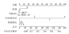
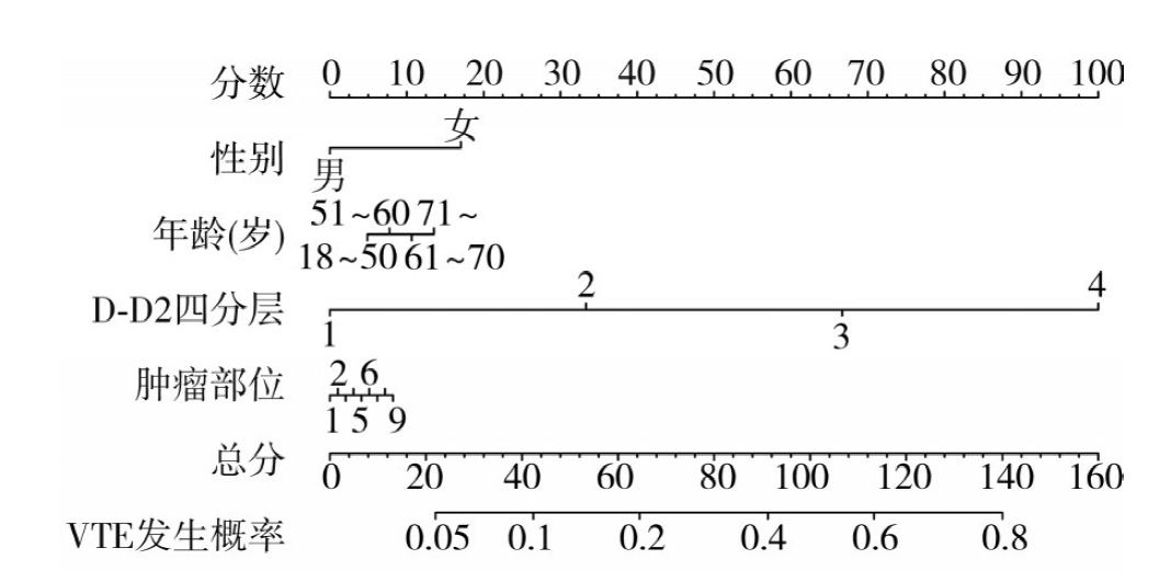
| 临床特征 | OR(95%CI) | P值 |
|---|---|---|
| 性别 | ||
| 男 | Ref | |
| 女 | 1.87(1.20~2.90) | 0.006 |
| 年龄(岁) | ||
| 18~50 | Ref | |
| 51~60 | 0.73(0.41~1.32) | 0.302 |
| 61~70 | 0.56(0.32~0.98) | 0.042 |
| 71~ | 0.64(0.35~1.16) | 0.141 |
| 肿瘤部位 | ||
| 头颈 | Ref | |
| 肝胆胰 | 0.60(0.20~1.81) | 0.364 |
| 肺/支气管 | 0.51(0.24~1.06) | 0.070 |
| 食管 | 0.30(0.14~0.67) | 0.003 |
| 胃肠 | 0.31(0.15~0.68) | 0.003 |
| 乳腺 | 0.15(0.07~0.33) | <0.001 |
| 妇科 | 1.05(0.49~2.21) | 0.907 |
| 泌尿 | 0.33(0.13~0.86) | 0.023 |
| 其他 | 0.71(0.28~1.79) | 0.468 |
| D-D1 (μg/L FEU) | ||
| ≤550 | Ref | |
| 551~1 100 | 0.86(0.53~1.39) | 0.530 |
| 1 101~4 000 | 0.90(0.56~1.43) | 0.651 |
| ≥4 001 | 1.26(0.72~2.20) | 0.420 |
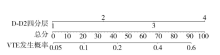
| D-D2 (μg/L FEU) | ||
| ≤550 | Ref | |
| 551~1 100 | 2.55(1.31~4.99) | 0.006 |
| 1 101~4 000 | 9.17(5.06~16.61) | <0.001 |
| ≥4 001 | 21.09(11.38~39.08) | <0.001 |
"
"


"
| [1] | Harry J, Bucciol R, Finnigan D, et al. The incidence of venous thromboembolism by type of solid cancer worldwide: a systematic review[J]. Cancer Epidemiol 2025, 95:102764. DOI: 10.1016/j.canep.2025.102764. |
| [2] | Bao Y, Wan X, Fu J, et al. The risk of venous thromboembolism in cancer patients receiving chemotherapy: a meta-analysis with systematic review[J]. Ann Transl Med, 2021, 9(4): 277. DOI: 10.21037/atm-20-3292. |
| [3] | 何进椅, 赵丽岩, 丁敏辉, 等. 静脉血栓栓塞症的流行病学与临床研究进展[J]. 中国心血管杂志, 2024, 29(6): 585-588. DOI: 10.3969/j.issn.1007-5410.2024.06.015. |
| [4] | Chen X, Huang J, Liu J, et al. Derivation and external validation of a risk assessment model of venous thromboembolism in hospitalized Chinese patients[J]. Clin Appl Thromb Hemost, 2023, 29: 10760296221151164. DOI: 10.1177/10760296221151164. |
| [5] | 孙维蔚, 姚学敏, 王鹏健, 等. 基于血液学指标探讨免疫治疗晚期非小细胞肺癌预后因素及列线图构建[J]. 国际肿瘤学杂志, 2024, 51(3): 143-150. DOI: 10.3760/cma.j.cn371439-20231109-00023. |
| [6] | Pabinger I, van Es N, Heinze G, et al. A clinical prediction model for cancer-associated venous thromboembolism: a development and validation study in two independent prospective cohorts[J]. Lancet Haematol, 2018, 5(7): e289-e298. DOI: 10.1016/S2352-3026(18)30063-2. |
| [7] | 钱伟伟, 徐申, 孔琪. 术前D二聚体在肾嗜酸细胞腺瘤和肾嫌色细胞癌中的鉴别诊断价值[J]. 国际肿瘤学杂志, 2023, 50(10): 614-617. DOI: 10.3760/cma.j.cn371439-20230612-00116. |
| [8] |
Hanna DL, White RH, Wun T. Biomolecular markers of cancer-associated thromboembolism[J]. Crit Rev Oncol Hematol, 2013, 88(1): 19-29. DOI: 10.1016/j.critrevonc.2013.02.008.
pmid: 23522921 |
| [9] |
Li W, Tang Y, Song Y, et al. Prognostic role of pretreatment plasma D-dimer in patients with solid tumors: a systematic review and meta-analysis[J]. Cell Physiol Biochem, 2018, 45(4): 1663-1676. DOI: 10.1159/000487734.
pmid: 29490291 |
| [10] | 中国研究型医院学会血栓与止血专业委员会. D-二聚体实验室检测与临床应用中国专家共识[J]. 中华医学杂志, 2023, 103(35): 2743-2756. DOI: 10.3760/cma.j.cn112137-20230721-00066. |
| [11] | Kench JG, Amin MB, Berney DM, et al. WHO classification of tumours fifth edition: evolving issues in the classification, diagnosis, and prognostication of prostate cancer[J]. Histopathology, 2022, 81(4): 447-458. DOI: 10.1111/his.14711. |
| [12] |
Obi AT, Pannucci CJ, Nackashi A, et al. Validation of the caprini venous thromboembolism risk assessment model in critically ill surgical patients[J]. JAMA Surg, 2015, 150(10): 941-948. DOI: 10.1001/jamasurg.2015.1841.
pmid: 26288124 |
| [13] | 何丽丽, 曹国磊, 罗琴. 凝血指标及肿瘤标志物与不同分期恶性肿瘤合并静脉血栓栓塞症的关系[J]. 中国医药导报, 2020, 17(17): 117-120. DOI: 10.20047/j.issn1673-7210.2020.17.028. |
| [14] | Horsted F, West J, Grainge MJ. Risk of venous thromboembolism in patients with cancer: a systematic review and meta-analysis[J]. PLoS Med, 2012, 9(7): e1001275. DOI: 10.1371/journal.pmed.1001275. |
| [15] |
Li J, Qiang WM, Wang Y, et al. Development and validation of a risk assessment nomogram for venous thromboembolism associated with hospitalized postoperative Chinese breast cancer patients[J]. J Adv Nurs, 2021, 77(1): 473-483. DOI: 10.1111/jan.14571.
pmid: 33159325 |
| [16] |
Timp JF, Braekkan SK, Versteeg HH, et al. Epidemiology of cancer-associated venous thrombosis[J]. Blood, 2013, 122(10): 1712-1723. DOI: 10.1182/blood-2013-04-460121.
pmid: 23908465 |
| [17] | 赵倩雯, 彭丹莉, 秦韬, 等. 1990—2019年全球肿瘤发病死亡分析[J]. 国际肿瘤学杂志, 2023, 50(7): 425-431. DOI: 10.3760/cma.j.cn371439-20230315-00082. |
| [18] | Abdol Razak NB, Jones G, Bhandari M, et al. Cancer-associated thrombosis: an overview of mechanisms, risk factors, and treatment[J]. Cancers (Basel), 2018, 10(10): 380. DOI: 10.3390/cancers10100380. |
| [19] | Alavi P, Rathod AM, Jahroudi N. Age-associated increase in thrombogenicity and its correlation with von willebrand factor[J]. J Clin Med, 2021, 10(18): 4190. DOI: 10.3390/jcm10184190. |
| [20] |
Ahlbrecht J, Dickmann B, Ay C, et al. Tumor grade is associated with venous thromboembolism in patients with cancer: results from the Vienna cancer and thrombosis study[J]. J Clin Oncol, 2012, 30(31): 3870-3875. DOI: 10.1200/JCO.2011.40.1810.
pmid: 23008313 |
| [21] | Mauriello A, Maratea AC, Fonderico C, et al. Physiopathological linkage and clinical perspectives[J]. Clin Med, 2025, 14(17): 6341. DOI: 10.3390/jcm14176341. |
| [22] | 《中国血栓性疾病防治指南》专家委员会. 中国血栓性疾病防治指南[J]. 中华医学杂志, 2018, 98(36): 2861-2888. DOI: 10.3760/cma.j.issn.0376-2491.2018.36.002. |
| [23] | 中华医学会呼吸病学分会肺栓塞与肺血管病学组, 中国医师协会呼吸医师分会肺栓塞与肺血管病工作委员会,全国肺栓塞与肺血管病防治协作组. 肺血栓栓塞症诊治与预防指南[J]. 中华医学杂志, 2018, 98(14): 1060-1087. DOI: 10.3760/cma.j.issn.0376-2491.2018.14.007. |
| [24] | 中华医学会外科学分会血管外科学组. 深静脉血栓形成的诊断和治疗指南(第三版)[J]. 中华普通外科杂志, 2017, 32(9): 807-812. DOI: 10.3760/cma.j.issn.1007-631X.2017.09.032. |
| [25] |
Ferroni P, Martini F, Portarena I, et al. Novel high-sensitive D-dimer determination predicts chemotherapy-associated venous thromboembolism in intermediate risk lung cancer patients[J]. Clin Lung Cancer, 2012, 13(6): 482-487. DOI: 10.1016/j.cllc.2012.03.005.
pmid: 22591606 |
| [26] |
Kodama J, Seki N, Masahiro S, et al. D-dimer level as a risk factor for postoperative venous thromboembolism in Japanese women with gynecologic cancer[J]. Ann Oncol, 2010, 21(8): 1651-1656. DOI: 10.1093/annonc/mdq012.
pmid: 20129998 |
| [27] |
Cohen AT, Spiro TE, Spyropoulos AC, et al. D-dimer as a predictor of venous thromboembolism in acutely ill, hospitalized patients: a subanalysis of the randomized controlled MAGELLAN trial[J]. J Thromb Haemost, 2014, 12(4): 479-487. DOI: 10.1111/jth.12515.
pmid: 24460645 |
| [28] | 王丽丽, 刘峰, 张燕捷, 等. 血浆D-二聚体水平与恶性肿瘤患者发生静脉血栓的相关性[J]. 现代肿瘤医学, 2018, 26(9): 1427-1431. DOI: 10.3969/j.issn.1672-4992.2018.09.029. |
| [29] | 陈青山, 章智荣, 董红红, 等. 《胸部恶性肿瘤围术期静脉血栓栓塞症预防中国专家共识(2018版)》解读之D-二聚体篇[J]. 中国肺癌杂志, 2019, 22(12): 761-766. DOI: 10.3779/j.issn.1009-3419.2019.12.05. |
| [30] | 赵雨婷, 张义静, 孙丽, 等. 肿瘤患者静脉血栓栓塞症风险评估的最佳证据总结[J]. 天津护理, 2024, 32(2): 193-197. DOI: 10.3969/j.issn.1006-9143.2024.02.015. |
| [31] |
Áinle FN, Kevane B. Which patients are at high risk of recurrent venous thromboembolism (deep vein thrombosis and pulmonary embolism)?[J]. Blood Adv, 2020, 4(21): 5595-5606. DOI: 10.1182/bloodadvances.2020002268.
pmid: 33170937 |
| Viewed | ||||||
|
Full text |
|
|||||
|
Abstract |
|
|||||