Journal of International Oncology ›› 2026, Vol. 53 ›› Issue (2): 65-72.doi: 10.3760/cma.j.cn371439-20250415-00010
• Original Article • Previous Articles Next Articles
Effects of cordycepin on proliferation, apoptosis and epithelial mesenchymal transition in nasopharyngeal carcinoma cells by regulating Akt/GSK-3β/Snail signaling pathway
Shi Rui1, Dai Jian2( ), Chen Ran1, Hu Lili1
- 1Department of Radiation Oncology, Cancer Center, Lu'an People's Hospital of Anhui Province, Lu'an 237000, China
2Department of Otolaryngology Head and Neck Surgery, Lu'an People's Hospital of Anhui Province, Lu'an 237000, China
-
Received:2025-04-15Online:2026-02-08Published:2026-01-29 -
Contact:Dai Jian E-mail:403619052@qq.com -
Supported by:Lu'an Municipal Scientific and Technological Program(2022lakj037)
Cite this article
Shi Rui, Dai Jian, Chen Ran, Hu Lili. Effects of cordycepin on proliferation, apoptosis and epithelial mesenchymal transition in nasopharyngeal carcinoma cells by regulating Akt/GSK-3β/Snail signaling pathway[J]. Journal of International Oncology, 2026, 53(2): 65-72.
share this article
"
| 虫草素浓度(μmol/L) | CNE-1细胞 | 5-8F细胞 | NP69细胞 |
|---|---|---|---|
| 0 | 98.16±3.95 | 98.89±4.18 | 97.27±3.86 |
| 1 | 93.43±3.67 | 97.26±4.02 | 96.02±3.79 |
| 2 | 89.58±3.57a | 95.12±3.86 | 96.59±3.81 |
| 5 | 83.94±3.26ab | 90.37±3.79ab | 94.64±3.26 |
| 10 | 71.85±3.01abcd | 79.58±3.54abcd | 93.78±3.56 |
| 20 | 62.72±2.84abcde | 67.64±3.37abcde | 94.35±3.17 |
| 40 | 50.79±2.61abcdef | 59.49±3.22abcdef | 93.71±3.25 |
| 80 | 37.73±2.72abcdefg | 51.13±3.01abcdefg | 93.19±3.42 |
| F值 | 269.03 | 154.81 | 1.08 |
| P值 | <0.001 | <0.001 | 0.393 |
"
"
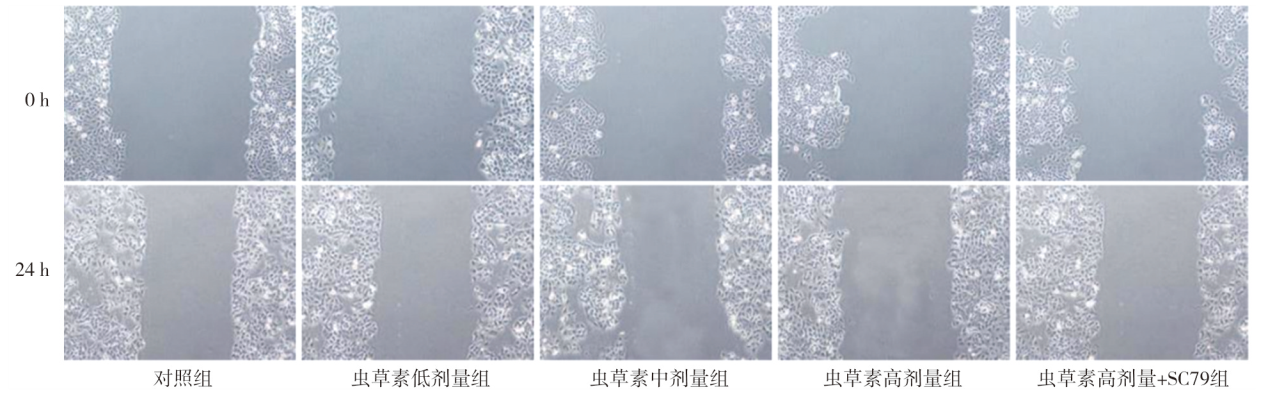
| 组别 | 细胞划痕愈合率(%) | 细胞侵袭个数(个) |
|---|---|---|
| 对照组 | 38.16±2.37 | 96.50±3.49 |
| 虫草素低剂量组 | 30.27±2.26a | 78.33±3.12a |
| 虫草素中剂量组 | 24.19±2.01ab | 62.17±2.93ab |
| 虫草素高剂量组 | 17.15±1.92abc | 43.50±2.58abc |
| 虫草素高剂量+SC79组 | 22.73±2.15d | 57.33±2.27d |
| F值 | 83.58 | 295.35 |
| P值 | <0.001 | <0.001 |
"
"
"
"
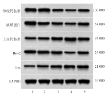
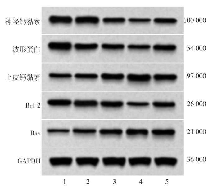
| 组别 | 神经钙黏素 | 波形蛋白 | 上皮钙黏素 | Bcl-2 | Bax |
|---|---|---|---|---|---|
| 对照组 | 0.82±0.08 | 0.94±0.09 | 0.42±0.04 | 0.89±0.08 | 0.31±0.03 |
| 虫草素低剂量组 | 0.68±0.05a | 0.76±0.07a | 0.57±0.06a | 0.73±0.06a | 0.45±0.05a |
| 虫草素中剂量组 | 0.55±0.04ab | 0.59±0.06ab | 0.73±0.07ab | 0.57±0.05ab | 0.61±0.06ab |
| 虫草素高剂量组 | 0.39±0.02abc | 0.42±0.03abc | 0.91±0.09abc | 0.43±0.03abc | 0.78±0.08abc |
| 虫草高剂量+SC79组 | 0.53±0.06d | 0.56±0.05d | 0.78±0.07d | 0.55±0.07d | 0.67±0.06d |
| F值 | 54.89 | 60.27 | 47.10 | 52.26 | 60.67 |
| P值 | <0.001 | <0.001 | <0.001 | <0.001 | <0.001 |
"
"
"
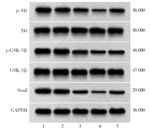
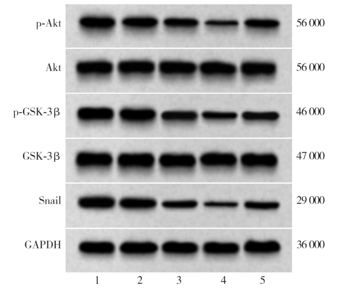
| 组别 | p-Akt/Akt | p-GSK-3β/GSK-3β | Snail |
|---|---|---|---|
| 对照组 | 0.87±0.08 | 0.98±0.09 | 0.92±0.08 |
| 虫草素低剂量组 | 0.75±0.06a | 0.84±0.08a | 0.74±0.05a |
| 虫草素中剂量组 | 0.60±0.05ab | 0.69±0.06ab | 0.56±0.05ab |
| 虫草素高剂量组 | 0.46±0.03abc | 0.54±0.05abc | 0.37±0.03abc |
| 虫草素高剂量+ SC79组 | 0.58±0.04d | 0.66±0.06d | 0.52±0.06d |
| F值 | 50.94 | 36.10 | 85.13 |
| P值 | <0.001 | <0.001 | <0.001 |
| [1] | He H, Liao X, Yang Q, et al. MicroRNA-494-3p promotes cell growth, migration, and invasion of nasopharyngeal carcinoma by targeting Sox7[J]. Technol Cancer Res Treat, 2018, 17: 1533033818809993. DOI: 10.1177/1533033818809993. |
| [2] | Liu YT, Hsieh MJ, Lin JT, et al. Erianin induces cell apoptosis through ERK pathway in human nasopharyngeal carcinoma[J]. Biomed Pharmacother, 2019, 111: 262-269. DOI: 10.1016/j.biopha.2018.12.081. |
| [3] | Zhang Z, Li K, Zheng Z, et al. Cordycepin inhibits colon cancer proliferation by suppressing MYC expression[J]. BMC Pharmacol Toxicol, 2022, 23(1): 12. DOI: 10.1186/s40360-022-00551-z. |
| [4] | Yoon SY, Park SJ, Park YJ. The anticancer properties of cordycepin and their underlying mechanisms[J]. Int J Mol Sci, 2018, 19(10): 3027. DOI: 10.3390/ijms19103027. |
| [5] | Zhou Y, Mei X, Li Y, et al. Cordycepin inhibits the proliferation and progression of NPC by targeting the MAPK/ERK and β-catenin pathways[J]. Oncol Lett, 2022, 23(1): 20. DOI: 10.3892/ol.2021.13138. |
| [6] | Lan Y, Han J, Wang Y, et al. STK17B promotes carcinogenesis and metastasis via AKT/GSK-3β/snail signaling in hepatocellular carcinoma[J]. Cell Death Dis, 2018, 9(2): 236. DOI: 10.1038/s41419-018-0262-1. |
| [7] | Wang N, Song Q, Yu H, et al. Overexpression of FBXO17 promotes the proliferation, migration and invasion of glioma cells through the Akt/GSK-3β/snail pathway[J]. Cell Transplant, 2021, 30: 9636897 211007395. DOI: 10.1177/09636897211007395. |
| [8] | 明华, 黄晓鸥, 林武华, 等. 山奈酚调节Akt/GSK-3β/Snail信号通路对鼻咽癌细胞增殖、凋亡和上皮间质转化的影响[J]. 现代肿瘤医学, 2024, 32(19): 3677-3683. DOI: 10.3969/j.issn.1672-4992.2024.19.009. |
| [9] | 李丹, 左淑萍, 张守军, 等. 虫草素改善糖尿病心肌病大鼠心肌组织及氧化应激作用的机制研究[J]. 中华老年心脑血管病杂志, 2025, 27(4): 504-509. DOI: 10.3969/j.issn.1009-0126.2025.04.023. |
| [10] |
Guo Z, Chen W, Dai G, et al. Cordycepin suppresses the migration and invasion of human liver cancer cells by downregulating the expression of CXCR4[J]. Int J Mol Med, 2020, 45(1): 141-150. DOI: 10.3892/ijmm.2019.4391.
pmid: 31746344 |
| [11] | 雷蕾, 高彦茹, 蔡洲, 等. 槐耳多糖通过抑制Akt/GSK3β/snail信号通路抑制胰腺癌细胞增殖和上皮间质转化[J]. 现代肿瘤医学, 2023, 31(24): 4536-4541. DOI: 10.3969/j.issn.1672-4992.2023.24.009. |
| [12] |
Chen C, Shan H. Keratin 6a gene silencing suppresses cell invasion and metastasis of nasopharyngeal carcinoma via the β‑catenin cascade[J]. Mol Med Rep, 2019, 19(5): 3477-3484. DOI: 10.3892/mmr.2019.10055.
pmid: 30896882 |
| [13] | 顾安琴, 龙金华, 金风. 鼻咽癌免疫治疗的临床研究进展[J]. 国际肿瘤学杂志, 2023, 50(5): 299-303. DOI: 10.3760/cma.j.cn371439-20230120-00060. |
| [14] | Wang Y, Jin W, Wang J. Tanshinone ⅡA regulates microRNA‑ 125b/foxp3/caspase‑1 signaling and inhibits cell viability of nasopharyngeal carcinoma[J]. Mol Med Rep, 2021, 23(5): 371. DOI: 10.3892/mmr.2021.12010. |
| [15] | Chen YY, Chen CH, Lin WC, et al. The role of autophagy in anti-cancer and health promoting effects of cordycepin[J]. Molecules, 2021, 26(16): 4954. DOI: 10.3390/molecules26164954. |
| [16] | Li HB, Chen JK, Su ZX, et al. Cordycepin augments the chemosensitivity of osteosarcoma to cisplatin by activating AMPK and suppressing the AKT signaling pathway[J]. Cancer Cell Int, 2021, 21(1): 706. DOI: 10.1186/s12935-021-02411-y. |
| [17] | Tung KL, Wu SZ, Yang CC, et al. Cordycepin induces apoptosis through JNK-mediated caspase activation in human OEC-M1 oral cancer cells[J]. Evid Based Complement Alternat Med, 2022, 2022(1): 1842363. DOI: 10.1155/2022/1842363. |
| [18] | 童汪霞, 罗宁, 李桂凤, 等. 虫草素通过调控Gli1介导抗肝癌细胞增殖及促凋亡的机制[J]. 中国实验方剂学杂志, 2022, 28(2): 104-111. DOI: 10.13422/j.cnki.syfjx.20220295. |
| [19] | Xu J, Shen X, Sun D, et al. Cordycepin suppresses the malignant phenotypes of colon cancer cells through the GSK3β/β-catenin/cyclin D1 signaling pathway[J]. Cell J, 2022, 24(5): 255-260. DOI: 10.22074/cellj.2022.8160. |
| [20] | Lorenzo-Herrero S, López-Soto A, Sordo-Bahamonde C, et al. NK cell-based immunotherapy in cancer metastasis[J]. Cancers (Basel), 2018, 11(1): 29. DOI: 10.3390/cancers11010029. |
| [21] |
Zhang K, Liu P, Tang H, et al. AFAP1-AS1 promotes epithelial-mesenchymal transition and tumorigenesis through Wnt/β-catenin signaling pathway in triple-negative breast cancer[J]. Front Pharmacol, 2018, 9: 1248. DOI: 10.3389/fphar.2018.01248.
pmid: 30505272 |
| [22] | Sun J, Chen W, Wen B, et al. Biejiajian pill inhibits carcinogenesis and metastasis via the Akt/GSK-3β/snail signaling pathway in hepatocellular carcinoma[J]. Front Pharmacol, 2021, 12: 610158. DOI: 10.3389/fphar.2021.610158. |
| [23] | Qiu WZ, Zhang HB, Xia WX, et al. The CXCL5/CXCR2 axis contributes to the epithelial-mesenchymal transition of nasopharyngeal carcinoma cells by activating ERK/GSK-3β/snail signalling[J]. J Exp Clin Cancer Res, 2018, 37(1): 85. DOI: 10.1186/s13046-018-0722-6. |
| [24] |
Yuan L, Zhou M, Huang D, et al. Resveratrol inhibits the invasion and metastasis of colon cancer through reversal of epithelial‑ mesenchymal transition via the Akt/GSK‑3β/snail signaling pathway[J]. Mol Med Rep, 2019, 20(3): 2783-2795. DOI: 10.3892/mmr.2019.10528.
pmid: 31524255 |
| [25] | Pan S, Liang S, Wang X. ADORA1 promotes nasopharyngeal carcinoma cell progression through regulation of PI3K/Akt/GSK-3β/β-catenin signaling[J]. Life Sci, 2021, 278: 119581. DOI: 10.1016/j.lfs.2021.119581. |
| Viewed | ||||||
|
Full text |
|
|||||
|
Abstract |
|
|||||