Journal of International Oncology ›› 2026, Vol. 53 ›› Issue (2): 73-78.doi: 10.3760/cma.j.cn371439-20250612-00011
• Original Article • Previous Articles Next Articles
Effect of SPART on the proliferation and migration capabilities of gastric cancer cell through lipophagy
Laibijiang Wusiman, Song Dingding, Zhang Wenbin( )
)
- Department of Gastrointestinal Surgery, Affiliated Tumor Hospital of Xinjiang Medical University, Urumqi 830000, China
-
Received:2025-06-12Online:2026-02-08Published:2026-01-29 -
Contact:Zhang Wenbin E-mail:zwb3216@sina.com -
Supported by:Xinjiang Uygur Autonomous Region Graduate Innovation Program(XJ2025G181);National Science Foundation of Xinjiang Uygur Autonomous Region of China(2022001D77)
Cite this article
Laibijiang Wusiman, Song Dingding, Zhang Wenbin. Effect of SPART on the proliferation and migration capabilities of gastric cancer cell through lipophagy[J]. Journal of International Oncology, 2026, 53(2): 73-78.
share this article

"
"
"
| 时间(h) | AGS细胞对照组 | AGS细胞实验组 | t值 | P值 | NUGC3细胞对照组 | NUGC3细胞实验组 | t值 | P值 |
|---|---|---|---|---|---|---|---|---|
| 0 | 0.20±0.01 | 0.20±0.01 | <0.01 | >0.999 | 0.33±0.01 | 0.33±0.01 | <0.01 | >0.999 |
| 24 | 0.29±0.01 | 0.30±0.01 | -1.22 | 0.290 | 0.46±0.01 | 0.45±0.01 | 1.22 | 0.290 |
| 48 | 0.52±0.01 | 0.41±0.01 | 13.47 | <0.001 | 0.67±0.02 | 0.51±0.01 | 12.39 | <0.001 |
| 72 | 0.82±0.01 | 0.67±0.01 | 18.37 | <0.001 | 1.05±0.01 | 0.78±0.02 | 20.91 | <0.001 |
| 96 | 1.04±0.03 | 0.74±0.01 | 16.43 | <0.001 | 1.80±0.02 | 1.41±0.08 | 8.19 | 0.004 |

"
"
"
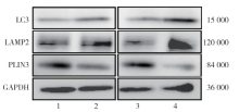
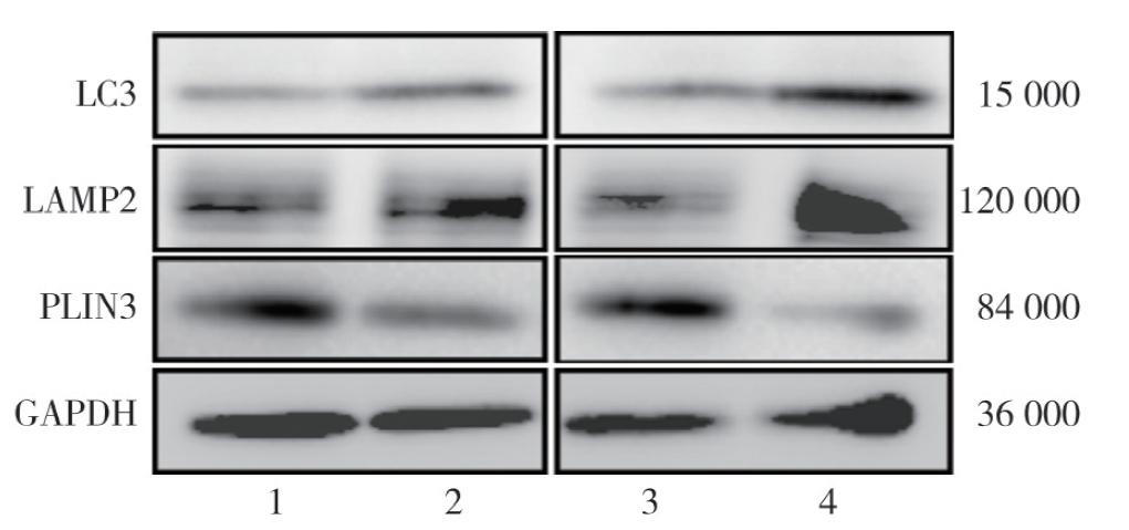
| 项目 | AGS细胞对照组 | AGS细胞实验组 | t值 | P值 | NUGC3细胞对照组 | NUGC3细胞实验组 | t值 | P值 |
|---|---|---|---|---|---|---|---|---|
| LC3 | 0.47±0.08 | 1.06±0.32 | 3.11 | 0.040 | 0.89±0.04 | 1.45±0.16 | 5.89 | 0.004 |
| LAMP2 | 0.65±0.03 | 0.98±0.11 | 5.02 | 0.007 | 0.68±0.18 | 1.22±0.22 | 3.29 | 0.030 |
| PLIN3 | 0.82±0.15 | 0.43±0.10 | 3.75 | 0.020 | 1.71±0.45 | 0.35±0.06 | 5.19 | 0.007 |
"
| [1] |
Sundar R, Nakayama I, Markar SR, et al. Gastric cancer[J]. Lancet, 2025, 405(10494): 2087-2102. DOI: 10.1016/S0140-6736(25)00052-2.
pmid: 40319897 |
| [2] | Siegel RL, Miller KD, Fuchs HE, et al. Cancer statistics, 2021[J]. CA Cancer J Clin, 2021, 71(1): 7-33. DOI: 10.3322/caac.21654. |
| [3] | Sung H, Ferlay J, Siegel RL, et al. Global cancer statistics 2020: GLOBOCAN estimates of incidence and mortality worldwide for 36 cancers in 185 countries[J]. CA Cancer J Clin, 2021, 71(3): 209-249. DOI: 10.3322/caac.21660. |
| [4] | Smyth EC, Verheij M, Allum W, et al. Gastric cancer: ESMO clinical practice guidelines for diagnosis, treatment and follow-up[J]. Ann Oncol, 2016, 27: v38-v49. DOI: 10.1093/annonc/mdw350. |
| [5] | Qiu H, Cao S, Xu R. Cancer incidence, mortality, and burden in China: a time-trend analysis and comparison with the United States and United Kingdom based on the global epidemiological data released in 2020[J]. Cancer Commun (Lond), 2021, 41(10): 1037-1048. DOI: 10.1002/cac2.12197. |
| [6] | Singh R, Kaushik S, Wang Y, et al. Autophagy regulates lipid metabolism[J]. Nature, 2009, 458(7242): 1131-1135. DOI: 10.1038/nature07976. |
| [7] | Cusenza VY, Bonora E, Amodio N, et al. Spartin: at the crossroad between ubiquitination and metabolism in cancer[J]. Biochim Biophys Acta Rev Cancer, 2022, 1877( 6): 188813. DOI: 10.1016/j.bbcan.2022.188813. |
| [8] |
Liu R, Lee JH, Li J, et al. Choline kinase alpha 2 acts as a protein kinase to promote lipolysis of lipid droplets[J]. Mol Cell, 2021, 81(13): 2722-2735.e9. DOI: 10.1016/j.molcel.2021.05.005.
pmid: 34077757 |
| [9] | 中国抗癌协会胃癌专业委员会,梁寒, 徐惠绵. 2024版CACA胃癌整合诊治指南(精简版)[J]. 中国肿瘤临床, 2024, 51(13): 650-657. DOI: 10.12354/j.issn.1000-8179.2024.20240882. |
| [10] |
Hanahan D. Hallmarks of cancer: new dimensions[J]. Cancer Discov, 2022, 12(1): 31-46. DOI: 10.1158/2159-8290.CD-21-1059.
pmid: 35022204 |
| [11] |
Kroemer G, Pouyssegur J. Tumor cell metabolism: cancer's Achilles' heel[J]. Cancer Cell, 2008, 13(6): 472-482. DOI: 10.1016/j.ccr.2008.05.005.
pmid: 18538731 |
| [12] | 谭荣坚, 欧雯婷, 翟嘉伟, 等. RRM2通过调控CDK1对胃癌细胞恶性生物学行为及有氧糖酵解的影响[J]. 国际肿瘤学杂志, 2025, 52(1): 23-30. DOI: 10.3760/cma.j.cn371439-20240607-00003. |
| [13] | 谭艳美, 谭艳飞, 蒙国照, 等. 脂噬在脂代谢疾病中的调控作用[J]. 中南医学科学杂志, 2022, 50(5): 777-780. DOI: 10.15972/j.cnki.43-1509/r.2022.05.039. |
| [14] | 王鑫, 宋海平, 王晔, 等. 细胞自噬与肿瘤的发生发展及治疗[J]. 国际肿瘤学杂志, 2018, 45(12): 743-746. DOI: 10.3760/cma.j.issn.1673-422X.2018.12.009. |
| [15] | Peng P, Chavel C, Liu W, et al. Pro-survival signaling regulates lipophagy essential for multiple myeloma resistance to stress-induced death[J]. Cell Rep, 2024, 43(7): 114445. DOI: 10.1016/j.celrep.2024.114445. |
| [16] | Liu S, Su Y, Han B, et al. Activation of Rab7-mediated lipophagy is required for triptolide to induce ferroptosis in hepatic cells[J]. Food Chem Toxicol, 2025, 203: 115568. DOI: 10.1016/j.fct.2025.115568. |
| [17] | Yin H, Shan Y, Xia T, et al. Emerging roles of lipophagy in cancer metastasis[J]. Cancers (Basel), 2022, 14(18): 4526. DOI: 10.3390/cancers14184526. |
| [18] |
Zhang M, Di Martino JS, Bowman RL, et al. Adipocyte-derived lipids mediate melanoma progression via FATP proteins[J]. Cancer Discov, 2018, 8(8): 1006-1025. DOI: 10.1158/2159-8290.CD-17-1371.
pmid: 29903879 |
| [19] |
Clark R, Krishnan V, Schoof M, et al. Milky spots promote ovarian cancer metastatic colonization of peritoneal adipose in experimental models[J]. Am J Pathol, 2013, 183(2): 576-591. DOI: 10.1016/j.ajpath.2013.04.023.
pmid: 23885715 |
| [20] | 孔秀枝, 单颖, 尤易文, 等. 雌激素相关受体α介导的脂噬对鼻咽癌细胞增殖和迁移能力的影响[J]. 肿瘤研究与临床, 2024, 36(2): 105-111. DOI: 10.3760/cma.j.cn115355-20230925-00123. |
| [21] | Geng F, Guo D. SREBF1/SREBP-1 concurrently regulates lipid synthesis and lipophagy to maintain lipid homeostasis and tumor growth[J]. Autophagy, 2024, 20(5): 1183-1185. DOI: 10.1080/15548627.2023.2275501. |
| [22] | Mukhopadhyay S, Schlaepfer IR, Bergman BC, et al. ATG14 facilitated lipophagy in cancer cells induce ER stress mediated mitoptosis through a ROS dependent pathway[J]. Free Radic Biol Med, 2017, 104: 199-213. DOI: 10.1016/j.freeradbiomed.2017.01.007. |
| [23] |
Pawella LM, Hashani M, Eiteneuer E, et al. Perilipin discerns chronic from acute hepatocellular steatosis[J]. J Hepatol, 2014, 60(3): 633-642. DOI: 10.1016/j.jhep.2013.11.007.
pmid: 24269473 |
| [24] |
Kaushik S, Cuervo AM. Degradation of lipid droplet-associated proteins by chaperone-mediated autophagy facilitates lipolysis[J]. Nat Cell Biol, 2015, 17(6): 759-770. DOI: 10.1038/ncb3166.
pmid: 25961502 |
| [25] | Tapia D, Jiménez T, Zamora C, et al. KDEL receptor regulates secretion by lysosome relocation-and autophagy-dependent modulation of lipid-droplet turnover[J]. Nat Commun, 2019, 10(1): 735. DOI: 10.1038/s41467-019-08501-w. |
| [26] | Filali-Mouncef Y, Hunter C, Roccio F, et al. The ménage à trois of autophagy, lipid droplets and liver disease[J]. Autophagy, 2022, 18(1): 50-72. DOI: 10.1080/15548627.2021.1895658 |
| [1] | Arya Ehmet, Nuriman Samat, Wang Tingting. Analysis of the correlation between nutritional status and preoperative gastric morphology and functional characteristics in patients with gastric cancer after radical gastrectomy [J]. Journal of International Oncology, 2026, 53(2): 87-92. |
| [2] | Yang Xinting, Ma Tengyu, Guan Shulong, Yang Mei, Jiang Zhou, Yang Xinru, Jiang Liangqian, Gao Meihua, Xu Yingjie, Cong Beibei. Research on the mechanism of targeting CD59 to inhibit proliferation, migration, and induce apoptosis in oral squamous cell carcinoma [J]. Journal of International Oncology, 2026, 53(1): 16-23. |
| [3] | Zhang Xiaoxi, Cheng Chunlai. Expression of ALDH6A1 in clear cell renal cell carcinoma and its impacts on proliferation, apoptosis, and invasion of renal cancer cells [J]. Journal of International Oncology, 2026, 53(1): 38-46. |
| [4] | Wang Yu, Li Yuanfei, Guo Yuntong. Research progress of the immunoscore system in gastric cancer [J]. Journal of International Oncology, 2026, 53(1): 62-64. |
| [5] | Liu Mei, Hu Yuchong, Li Fengtong, Chao Lemen, Liu Meng, Kang Linlin. Mechanism of action of SHCBP1 in malignant tumors and progress in clinical research [J]. Journal of International Oncology, 2025, 52(9): 583-586. |
| [6] | Li Guangxin, Quan Huijuan, Gao Zhijuan, Wang Xiaojun, Li Liang, Dong Qian, Miao Yongtao, Liu Dongsheng. Correlation between serum levels of HAMP, SPP1, RGS2 and clinical pathological characteristics of gastric cancer patients and their predictive value for postoperative recurrence or metastasis [J]. Journal of International Oncology, 2025, 52(8): 502-507. |
| [7] | Liu Shan, Pan Yue, Zhang Zhuo, Liu Chong, Li Xueman, Xiong Fei. Inhibition of the growth, migration, and angiogenesis of esophageal squamous cell carcinoma by metformin by regulating ALKBH3 expression [J]. Journal of International Oncology, 2025, 52(6): 343-352. |
| [8] | Liu Qianyi, Dong Hongmin, Wang Wenling, Wang Gang, Chen Wanghua. Clinical efficacy and safety of radiotherapy combined with chemotherapy and immunotherapy for HER2-negative locally advanced or advanced gastric cancer [J]. Journal of International Oncology, 2025, 52(4): 209-216. |
| [9] | Ji Haitao, Wang Yanfeng, Liu Yongcheng, Hao Nan. Expression and clinical significance of DHCR7 in gastric cancer based on bioinformatics analysis [J]. Journal of International Oncology, 2025, 52(2): 94-100. |
| [10] | Liu Pingping, Wang Junyi, Lin Zhiwei, Chen Dachao. Analysis of factors influencing the prognosis of patients with postoperative peritoneal metastasis of gastric cancer [J]. Journal of International Oncology, 2025, 52(12): 764-769. |
| [11] | Zhang Shuai, Liu Liangliang, Huang Di, Sheng Ru, Qi Mengyao, Li Shuguang. Effects of circRNA-15430 targeting miR-10 on proliferation and invasion of gastric cancer cells [J]. Journal of International Oncology, 2025, 52(11): 673-679. |
| [12] | Tan Rongjian, Ou Wenting, Zhai Jiawei, Quan Zhenhao, Sun Lijun, Zhou Caijin. Effects of RRM2 on malignant biological behavior and aerobic glycolysis of gastric cancer cells by regulating CDK1 [J]. Journal of International Oncology, 2025, 52(1): 23-30. |
| [13] | Wu Yang, Li Tian, Zhang Runbing, Shi Tingting, Gao Chun, Zheng Xiaofeng, Zhang Jiucong. Research progress in immunotherapy and targeted therapy for gastric cancer and esophagogastric junction cancer [J]. Journal of International Oncology, 2024, 51(9): 595-600. |
| [14] | Liu Wenhui, Yin Ping, Qi Jie. Diagnostic value of detection of serum G-17,sB7-H3,and DKK1 for early gastric cancer [J]. Journal of International Oncology, 2024, 51(8): 498-503. |
| [15] | Yuan Jian, Huang Yanhua. Diagnostic value of Hp-IgG antibody combined with serum DKK1 and sB7-H3 in early gastric cancer [J]. Journal of International Oncology, 2024, 51(6): 338-343. |
| Viewed | ||||||
|
Full text |
|
|||||
|
Abstract |
|
|||||