
Journal of International Oncology ›› 2025, Vol. 52 ›› Issue (9): 554-559.doi: 10.3760/cma.j.cn371439-20250530-00094
• Original Article • Previous Articles Next Articles
Li Peng1, Zhang Shuang1, Liu Huafeng2, Ji Na2, Hou Xiangkun2, Xi Aohang2, Zong Jianhai2( )
)
Received:2025-05-30
Revised:2025-07-20
Online:2025-09-08
Published:2025-10-21
Contact:
Zong Jianhai
E-mail:1911688816@qq.com
Li Peng, Zhang Shuang, Liu Huafeng, Ji Na, Hou Xiangkun, Xi Aohang, Zong Jianhai. Research on positioning errors analysis of gamma knife pain-free face mask fractionated treatment for head tumors based on kV orthogonal image guidance[J]. Journal of International Oncology, 2025, 52(9): 554-559.
"
"
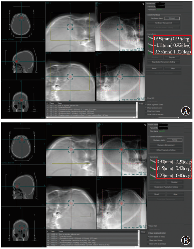
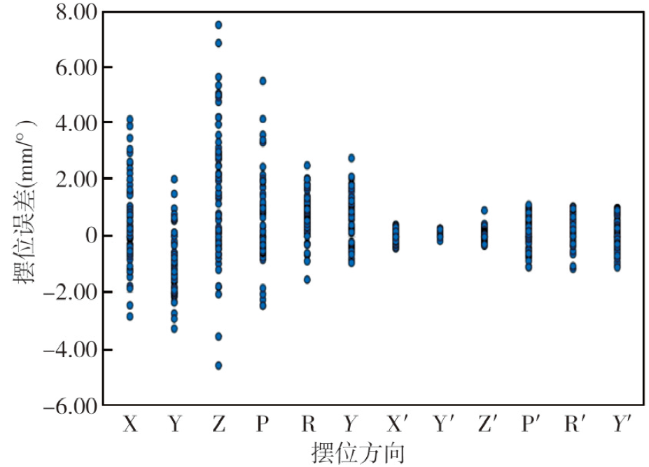
| 项目 | X (mm) | Y(mm) | Z(mm) | P(°) | R(°) | Y(°) |
|---|---|---|---|---|---|---|
| 校正前 | 0.45±1.54 | -0.96(-1.70,-0.28) | 1.67(-0.15,3.07) | 0.70±1.60 | 0.65(0.30,1.19) | 0.59±0.87 |
| 校正后 | -0.02±0.18 | 0.15(0.10,0.21) | 0.06(-0.04,0.16) | 0.20±0.79 | 0.42(0.19,0.78) | 0.20±0.63 |
| t/Z值 | 2.30 | -5.43 | -4.10 | 2.56 | -3.21 | 3.21 |
| P值 | 0.025 | <0.001 | <0.001 | 0.013 | 0.001 | 0.002 |

"
"
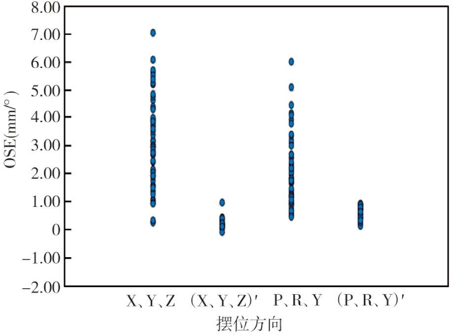
| 项目 | 平移OSE(X、Y、Z)(mm) | 旋转OSE(P、R、Y)(°) |
|---|---|---|
| 校正前 | 3.07(1.93,4.35) | 1.90(1.28,2.66) |
| 校正后 | 0.27(0.21,0.33) | 1.08(0.70,1.54) |
| Z值 | -6.60 | -5.52 |
| P值 | <0.001 | <0.001 |

"
"
| 年龄段(岁) | 校正前OSE(X、Y、Z)(mm) | 校正后OSE(X、Y、Z)(mm) | 校正前OSE(P、R、Y)(°) | 校正后OSE(P、R、Y)(°) |
|---|---|---|---|---|
| 18~44(n=7) | 3.65(1.62,3.95) | 0.21(0.21,0.31) | 3.25(2.24,3.96) | 0.92(0.59,1.45) |
| 45~59(n=19) | 3.57(2.17,5.22) | 0.29(0.22,0.35) | 1.89(1.30,2.30) | 1.08(0.62,1.51) |
| 60~74(n=25) | 2.92(1.74,4.06) | 0.24(0.19,0.35) | 2.16(1.09,2.95) | 0.98(0.78,1.75) |
| 75~89(n=7) | 3.24(2.12,4.37) | 0.29(0.22,0.47) | 1.73(1.01,1.83) | 0.60(0.47,1.51) |
| H值 | 1.23 | 1.74 | 7.45 | 2.80 |
| P值 | 0.747 | 0.627 | 0.059 | 0.424 |
"
| 性别 | 校正前OSE(X、Y、Z)(mm) | 校正后OSE(X、Y、Z)(mm) | 校正前OSE(P、R、Y)(º) | 校正后OSE(P、R、Y)(º) |
|---|---|---|---|---|
| 男(n=33) | 3.19±1.59 | 0.27(0.27,0.33) | 1.89(1.27,2.75) | 0.84±0.59 |
| 女(n=25) | 3.22±1.99 | 0.26(0.25,0.35) | 1.90(1.34,2.41) | 1.04±0.46 |
| t/Z值 | -0.07 | -0.48 | -0.02 | -2.80 |
| P值 | 0.949 | 0.632 | 0.161 | 0.424 |
| [1] |
Nguyen T, Hsu W, Lim M, et al. Delivery of stereotactic radiosurgery: a cross-platform comparison[J]. Neurol Res, 2011, 33(8): 787-791. DOI: 10.1179/016164111X13123658647409.
pmid: 22004701 |
| [2] | Agrawal M, Mishra S, Garg K, et al. Trends in stereotactic radiosurgery for intracranial and spinal pathologies: analysis of the top 100 most cited articles[J]. Neurol India, 2023, 71 (Supplement): S39-S48. DOI: 10.4103/0028-3886.373651. |
| [3] | Tripathi M, Jani P, Bhatta R, et al. Development, validation, and impact of patient information booklet for gamma knife radiosurgery[J]. Neurol India, 2023, 71 (Supplement): S224-S229. DOI: 10.4103/0028-3886.373624. |
| [4] |
Inserra F, Barone F, Palmisciano P, et al. Hypofractionated gamma knife radiosurgery: institutional experience on benign and malignant intracranial tumors[J]. Anticancer Res, 2022, 42(4): 1851-1858. DOI: 10.21873/anticanres.15661.
pmid: 35347003 |
| [5] | Tripathi M, Chauhan R, Luthra A, et al. Anesthetic concerns during gamma-knife radiosurgery[J]. Neurol India, 2023, 71(Supplement): S74-S81. DOI: 10.4103/0028-3886.373626. |
| [6] | Yamaguchi H. Gamma knife radiosurgery with mask fixation under general anesthesia for pediatric patients[J]. Cureus, 2022, 14(1): 20905. DOI: 10.7759/cureus.20905. |
| [7] |
Grimm MA, Köppen U, Stieler F, et al. Prospective assessment of mask versus frame fixation during gamma knife treatment for brain metastases[J]. Radiother Oncol, 2020, 147: 195-199. DOI: 10.1016/j.radonc.2020.05.011.
pmid: 32416280 |
| [8] |
Claps L, Mathew D, Dusenbery K, et al. Utilization of CBCT to improve the delivery accuracy of gamma knife radiosurgery with G-frame[J]. J Appl Clin Med Phys, 2021, 22(8): 120-128. DOI: 10.1002/acm2.13332.
pmid: 34196098 |
| [9] | Caudell JJ, Gillison ML, Maghami E, et al. NCCN guidelines® insights: head and neck cancers, version 1.2022[J]. J Natl Compr Canc Netw, 2022, 20(3): 224-234. DOI: 10.6004/jnccn.2022.0016. |
| [10] | Chew JJ, Sneed PK, Chang EF. Recurrent radiation-induced cavernous malformation after gamma knife stereotactic radiosurgery for brain metastasis[J]. Cureus, 2022, 14(3): e22815. DOI: 10.7759/cureus.22815. |
| [11] | Moon HC, Park SJ, Kim YD, et al. Navigation of frameless fixation for gamma knife radiosurgery using fixed augmented reality[J]. Sci Rep, 2022, 12(1): 4486. DOI: 10.1038/s41598-022-08390-y. |
| [12] | Li Z, Cheng Y, Dong J, et al. The impact of setup errors on dose distribution in cervical cancer radiotherapy and the margin from CTV to PTV[J]. J Cancer Res Clin Oncol, 2024, 150(12): 516. DOI: 10.1007/s00432-024-06032-6. |
| [13] | Luo G, Neimat JS, Cmelak A, et al. Margin of error for a frameless image guided radiosurgery system: direct confirmation based on posttreatment MRI scans[J]. Pract Radiat Oncol, 2017, 7(3): e223-e231. DOI: 10.1016/j.prro.2016.08.006. |
| [14] | 张彦新, 符贵山, 徐英杰, 等. 脑转移瘤立体定向放疗分次间和分次内摆位误差及残余误差分析[J]. 中华放射肿瘤学杂志, 2019, 28(6): 448-451. DOI: 10.3760/cma.j.issn.1004-4221.2019.06.012. |
| [15] | 张平, 罗龙辉, 戴鹏, 等. ExacTrac 6D影像精确引导系统在脑转移瘤SRS中的应用[J]. 中国医疗设备, 2017, 32(5): 40-43. DOI: 10.3969/j.issn.1674-1633.2017.05.010. |
| [16] |
Tanaka Y, Oita M, Inomata S, et al. Impact of patient positioning uncertainty in noncoplanar intracranial stereotactic radiotherapy[J]. J Appl Clin Med Phys, 2020, 21(2): 89-97. DOI: 10.1002/acm2.12820.
pmid: 31957975 |
| [17] |
Faraj MK, Alazawy NM, Madlool SK, et al. Navigating precision: anatomical insights into the efficacy of masks versus stereotactic frames in icon gamma knife treatment for trigeminal neuralgia[J]. Surg Neurol Int, 2025, 16: 3. DOI: 10.25259/SNI_757_2024.
pmid: 39926454 |
| [18] | Gill M, Sharma M, Ratan R. Frameless gamma knife radiosurgery with leksell ICON: initial experience[J]. Neurol India, 2023, 71 (Supplement): S68-S73. DOI: 10.4103/0028-3886.373646. |
| [19] | Faraj MK, Al-Musawi MS, Ali Abdulameer T. Design and manufacturing of a head mask for fixation in stereotactic radiosurgery by the gamma knife icon[J]. Surg Neurol Int, 2023, 14: 188. DOI: 10.25259/SNI_1053_2022. |
| [1] | Chen Qi, Xu Chenyang, Wang Yin, Lei Dapeng. Current application status of hyperspectral imaging in the diagnosis and treatment of head and neck tumor [J]. Journal of International Oncology, 2024, 51(5): 298-302. |
| [2] | Cui Tenglu, Lyu lu, Sun Pengfei. Application of radiotherapy combined with immunotherapy in the treatment of head and neck squamous cell carcinoma [J]. Journal of International Oncology, 2023, 50(9): 548-552. |
| [3] | Chen Xinyi, Weng Yiming, Wei Jiayan, Wang Jinsong, Peng Min. Advances in immune checkpoint inhibitors in the treatment of recurrent or metastatic head and neck squamous cell carcinoma [J]. Journal of International Oncology, 2023, 50(9): 553-557. |
| [4] | Ju Yifan, Xu Chenyang, Lei Dapeng. Research progress of pathomics in head and neck neoplasms [J]. Journal of International Oncology, 2023, 50(5): 294-298. |
| [5] | Zhao Yongrui, Gao Ying, Chen Yidong, Xu Jiankun. Efficacy and safety of linear accelerator-based fractionated stereotactic radiotherapy for small volume brain metastases [J]. Journal of International Oncology, 2023, 50(3): 138-143. |
| [6] | Chen Yi, Han Liang, Cai Li. Multivariate analysis of chemotherapy induced oral mucositis in patients with head and neck tumors [J]. Journal of International Oncology, 2022, 49(9): 521-525. |
| [7] | Zeng Yan, Luo Pan, Wang Ziqi, Wu Weili. Mechanism of drug induced ferroptosis in the treatment of head and neck tumors [J]. Journal of International Oncology, 2022, 49(3): 173-176. |
| [8] | Lao Zheng, Tu Wenyong, Xu Xuanli, Zhang Lin, Shao Ziyang, Shi Huifeng. Nimotuzumab combined with definitive radiotherapy for inoperable locally advanced oral and maxillofacial squamous cell carcinoma [J]. Journal of International Oncology, 2022, 49(11): 665-670. |
| [9] | Du Xiao, Zhou Juying. Stereotactic body radiotherapy for localized prostate cancer [J]. Journal of International Oncology, 2021, 48(5): 313-316. |
| [10] | Xu Shifei, Feng Huan, Liu Haiyang, Hu Jie, Ma Lu. Effect of rotational errors on the accuracy of positioning for head-neck tumors in radiotherapy [J]. Journal of International Oncology, 2021, 48(3): 150-155. |
| [11] | Qian Wenchuan, Wang Fan. A dosimetric comparison of volumetric modulated arc therapy with intensity modulated radiation therapy for head and neck cancer [J]. Journal of International Oncology, 2020, 47(8): 457-461. |
| [12] | Zhou Fei, Liu Rui, Lyu Hongying, Liang Donghai, Chen Wenxiu, Yu Hongsheng. Immunotherapy for head and neck cancer [J]. Journal of International Oncology, 2020, 47(12): 746-751. |
| [13] | Chai Yue, Dong Mei . Medical treatment in head and neck squamous cell carcinoma [J]. Journal of International Oncology, 2019, 46(2): 94-97. |
| [14] | Zang Shoumei, Yan Danfang, Yan Senxiang. Advances in nonsurgical treatment of head and neck squamous cell carcinoma [J]. Journal of International Oncology, 2019, 46(12): 741-744. |
| [15] | DU Wei-Guang. Curative effect of stereotactic radiotherapy combined with TACE for patients with hepatic metastases of colorectal cancer and influence on the expressions of omentin-1 and Ang-2 [J]. Journal of International Oncology, 2018, 45(2): 87-91. |
| Viewed | ||||||
|
Full text |
|
|||||
|
Abstract |
|
|||||