Journal of International Oncology ›› 2025, Vol. 52 ›› Issue (9): 545-553.doi: 10.3760/cma.j.cn371439-20250704-00093
• Original Article • Previous Articles Next Articles
Anti-tumor effect and immunomodulatory mechanism of atractylenolide Ⅱ on colon cancer mice
Wang Mengju, Wang Xia( )
)
- Department of Oncology, Jiangsu Second Chinese Medicine Hospital, Second Affiliated Hospital of Nanjing University of Chinese Medicine, Nanjing 210017, China
-
Received:2025-07-04Revised:2025-07-19Online:2025-09-08Published:2025-10-21 -
Contact:Wang Xia E-mail:wangxia7904@163.com -
Supported by:National Natural Science Foundation of China(82474375);Hospital-Level Project of Jiangsu Second Chinese Medicine Hospital, Second Affiliated Hospital of Nanjing University of Chinese Medicine(SEZJY2023015)
Cite this article
Wang Mengju, Wang Xia. Anti-tumor effect and immunomodulatory mechanism of atractylenolide Ⅱ on colon cancer mice[J]. Journal of International Oncology, 2025, 52(9): 545-553.
share this article
"
| 组别 | 体质量(g) | 肿瘤体积(mm3) | 肿瘤质量(g) | 脾脏指数 |
|---|---|---|---|---|
| 模型组 | 20.54±1.07 | 1 845.17±65.72 | 1.34±0.11 | 7.42±0.88 |
| ATL-Ⅱ低剂量组 | 20.08±0.88 | 1 637.20±122.65a | 1.26±0.09 | 7.38±1.32 |
| ATL-Ⅱ中剂量组 | 20.12±1.63 | 1 232.86±209.16ab | 0.93±0.07ab | 8.42±0.78ab |
| ATL-Ⅱ高剂量组 | 20.46±1.87 | 1 002.29±41.84abc | 0.94±0.10ab | 9.72±1.18abc |
| 5-FU组 | 19.00±1.60 | 911.59±294.71abcd | 0.59±0.08abcd | 6.16±1.05abcd |
| F值 | 1.96 | 125.61 | 88.88 | 33.20 |
| P值 | 0.451 | <0.001 | <0.001 | <0.001 |
"
"
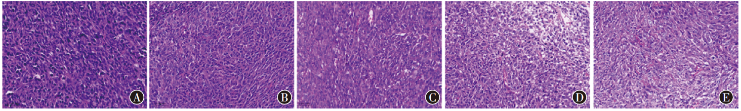
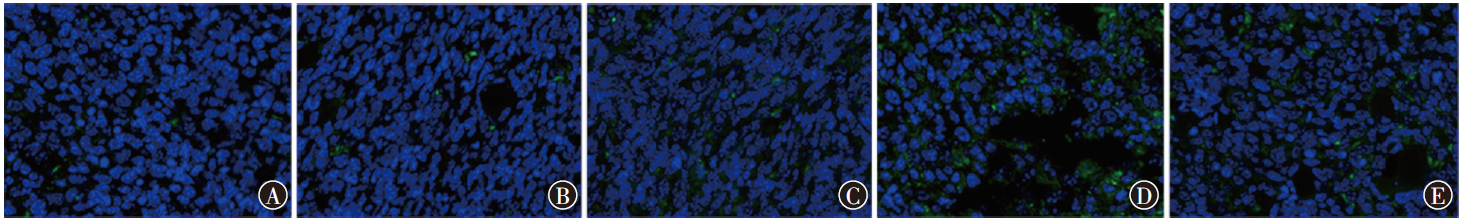
| 组别 | Ki-67 | Caspase-3 | Bcl-2 |
|---|---|---|---|
| 模型组 | 32.33±9.50 | 6.67±1.53 | 93.00±6.56 |
| ATL-Ⅱ低剂量组 | 24.33±5.69 | 11.00±2.65 | 94.00±12.12 |
| ATL-Ⅱ中剂量组 | 19.33±2.52a | 16.33±3.06a | 51.67±5.51ab |
| ATL-Ⅱ高剂量组 | 18.67±4.73ab | 52.33±4.04abc | 47.67±4.51ab |
| 5-FU组 | 16.33±2.52ab | 65.67±3.51abcd | 40.33±3.21ab |
| F值 | 13.86 | 477.63 | 40.48 |
| P值 | 0.043 | <0.001 | <0.001 |
"

"

"
"
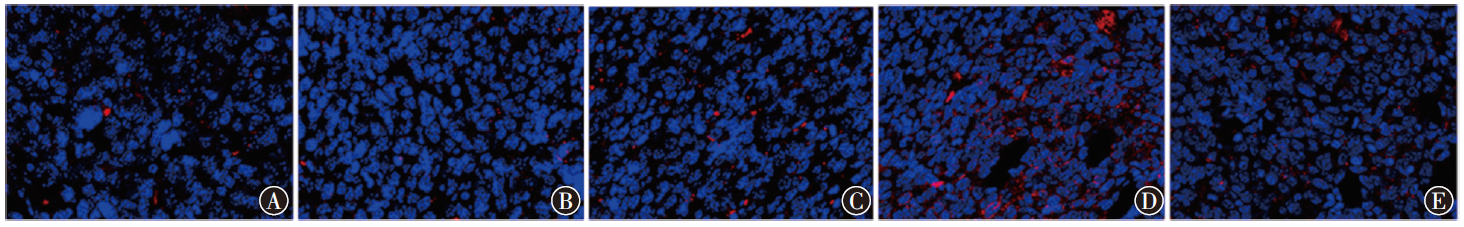
| 组别 | GzmB | IFN-λ |
|---|---|---|
| 模型组 | 5.00±1.00 | 617.33±65.06 |
| ATL-Ⅱ低剂量组 | 5.27±0.76 | 743.33±40.41a |
| ATL-Ⅱ中剂量组 | 8.27±0.61ab | 910.00±36.06ab |
| ATL-Ⅱ高剂量组 | 10.00±1.21abc | 1 009.00±35.54abc |
| 5-FU组 | 6.15±0.69acd | 703.62±56.00cd |
| F值 | 21.45 | 43.08 |
| P值 | <0.001 | <0.001 |
"
| 组别 | PD-L1 | p-ERK/ERK | p-MEK/MEK |
|---|---|---|---|
| 模型组 | 1.58±0.25 | 1.33±0.20 | 1.04±0.39 |
| ATL-Ⅱ低剂量组 | 0.88±0.24a | 0.61±0.21a | 0.74±0.24a |
| ATL-Ⅱ中剂量组 | 0.91±0.18a | 0.44±0.29ab | 0.70±0.20a |
| ATL-Ⅱ高剂量组 | 0.62±0.11abc | 0.57±0.15ab | 0.71±0.17a |
| 5-FU组 | 0.54±0.17abc | 0.23±0.11abcd | 0.63±0.19ab |
| F值 | 125.34 | 89.63 | 35.33 |
| P值 | <0.001 | <0.001 | 0.002 |
| [1] | Abedizadeh R, Majidi F, Khorasani HR, et al. Colorectal cancer: a comprehensive review of carcinogenesis, diagnosis, and novel strategies for classified treatments[J]. Cancer Metastasis Rev, 2024, 43(2): 729-753. DOI: 10.1007/s10555-023-10158-3. |
| [2] |
Fan A, Wang B, Wang X, et al. Immunotherapy in colorectal cancer: current achievements and future perspective[J]. Int J Biol Sci, 2021, 17(14): 3837-3849. DOI: 10.7150/ijbs.64077.
pmid: 34671202 |
| [3] | Lin Y, Chen K, Zhu M, et al. Atractylenolide Ⅱ regulates the proli-feration, ferroptosis, and immune escape of hepatocellular carcinoma cells by inactivating the TRAF6/NF-κB pathway[J]. Naunyn Schmiedebergs Arch Pharmacol, 2024, 397(10): 7697-7710. DOI: 10.1007/s00210-024-03046-2. |
| [4] | Tian S, Ren L, Liu C, et al. Atractylenolide Ⅱ suppresses glycolysis and induces apoptosis by blocking the PADI3-ERK signaling pathway in endometrial cancer cells[J]. Molecules, 2024, 29(5): 939. DOI: 10.3390/molecules29050939. |
| [5] | Zhang Y, Liu Y, Wang J, et al. Atractylenolide Ⅱ inhibits tumor-associated macrophages (TAMs)-induced lung cancer cell metastasis[J]. Immunopharmacol Immunotoxicol, 2022, 44(2): 227-237. DOI: 10.1080/08923973.2022.2037629. |
| [6] |
Lei X, Lei Y, Li JK, et al. Immune cells within the tumor micro-environment: biological functions and roles in cancer immunotherapy[J]. Cancer Lett, 2020, 470: 126-133. DOI: 10.1016/j.canlet.2019.11.009.
pmid: 31730903 |
| [7] | Zhang R, Wang Z, Yu Q, et al. Atractylenolide Ⅱ reverses the influence of lncRNA XIST/miR-30a-5p/ROR1 axis on chemo-resistance of colorectal cancer cells[J]. J Cell Mol Med, 2019, 23(5): 3151-3165. DOI: 10.1111/jcmm.14148. |
| [8] | Xie Z, Lin M, He X, et al. Chemical constitution, pharmacological effects and the underlying mechanism of atractylenolides: a review[J]. Molecules, 2023, 28(10): 3987. DOI: 10.3390/molecules28103987. |
| [9] | Wu Y, Dai S, Zhang Y, et al. Atractylenolide Ⅱcombined with interferon-γ synergistically ameliorates colorectal cancer progression in vivo and in vitro by blocking the NF-κB p65/PD-L1 pathway[J]. J Cancer, 2024, 15(13): 4328-4344. DOI: 10.7150/jca.96647. |
| [10] |
Sahoo G, Samal D, Khandayataray P, et al. A review on caspases: key regulators of biological activities and apoptosis[J]. Mol Neurobiol, 2023, 60(10): 5805-5837. DOI: 10.1007/s12035-023-03433-5.
pmid: 37349620 |
| [11] | Wang J, Nasser MI, Adlat S, et al. Atractylenolide Ⅱ induces apoptosis of prostate cancer cells through regulation of AR and JAK2/STAT3 signaling pathways[J]. Molecules, 2018, 23(12): 3298. DOI: 10.3390/molecules23123298. |
| [12] | Shang S, Yang YW, Chen F, et al. TRIB3 reduces CD8+ T cell infiltration and induces immune evasion by repressing the STAT1-CXCL10 axis in colorectal cancer[J]. Sci Transl Med, 2022, 14(626): eabf0992. DOI: 10.1126/scitranslmed.abf0992. |
| [13] |
Basu R, Whitlock BM, Husson J, et al. Cytotoxic T cells use mechanical force to potentiate target cell killing[J]. Cell, 2016, 165(1): 100-110. DOI: 10.1016/j.cell.2016.01.021.
pmid: 26924577 |
| [14] | Remsik J, Tong X, Kunes RZ, et al. Interferon-γ orchestrates leptomeningeal anti-tumour response[J]. Nature, 2025, 643(8073): 1087-1096. DOI: 10.1038/s41586-025-09012-z. |
| [15] | Zhou Y, Cheng L, Liu L, et al. NK cells are never alone: crosstalk and communication in tumour microenvironments[J]. Mol Cancer, 2023, 22(1): 34. DOI: 10.1186/s12943-023-01737-7. |
| [16] |
Huntington ND, Cursons J, Rautela J. The cancer-natural killer cell immunity cycle[J]. Nat Rev Cancer, 2020, 20(8): 437-454. DOI: 10.1038/s41568-020-0272-z.
pmid: 32581320 |
| [17] | Zheng Z, Wieder T, Mauerer B, et al. T cells in colorectal cancer: unravelling the function of different T cell subsets in the tumor microenvironment[J]. Int J Mol Sci, 2023, 24(14): 11673. DOI: 10.3390/ijms241411673. |
| [18] |
Fang JY, Richardson BC. The MAPK signalling pathways and colorectal cancer[J]. Lancet Oncol, 2005, 6(5): 322-327. DOI: 10.1016/S1470-2045(05)70168-6.
pmid: 15863380 |
| [19] | Wang L, Wang J, Wang N, et al. ANLN promotes head and neck squamous cell carcinoma progression by upregulating PD-L1 via the ERK-MAPK pathway[J]. iScience, 2025, 28(2): 111633. DOI: 10.1016/j.isci.2024.111633. |
| [1] | Liu Yiyong, Huo Fengzhi. Differential diagnosis model construction of invasive degree of lung adenocarcinoma manifesting as ground-glass nodules with no or little solid component based on energy spectrum CT features [J]. Journal of International Oncology, 2025, 52(4): 197-201. |
| [2] | Wei Wei, Cai Zhaoying, Qian Yayun. Effect of Marsdenia tenacissima combined with XELOX solution on disulfide apoptosis in human colorectal cancer HCT116 cells [J]. Journal of International Oncology, 2024, 51(9): 545-555. |
| [3] | Ren Lu, Xie Xiaoli, Zhang Kun, Wang Lijuan. Effects and mechanisms of dihydroartemisinin combined with carfilzomib on the activity, proliferation, and apoptosis of multiple myeloma cells [J]. Journal of International Oncology, 2024, 51(3): 129-136. |
| [4] | Xue Xiaofang, Zhong Yuquan, Li Xinyang. Construction of a prognostic model of hepatocellular carcinoma associated with lncRNA AL445524.1 and its effect on the malignant phenotype of hepatocellular carcinoma cells [J]. Journal of International Oncology, 2024, 51(11): 696-705. |
| [5] | Zhang Keping, Zhao Yongsheng, Yang Juan, Fu Maoyong. Chlorogenic acid induces mitochondrial dysfunction in lung cancer A549 cells by inhibiting the PI3K-Akt pathway [J]. Journal of International Oncology, 2024, 51(1): 21-28. |
| [6] | Xiang Yuling, Tan Jiajie, Xiong Yuanguo, Zhao Lirong, Li Chen, Zhang Hong. Effects of Anhydroicaritin on the proliferation, migration and apoptosis of hepatocellular carcinoma cells [J]. Journal of International Oncology, 2023, 50(9): 513-519. |
| [7] | Feng Dongxu, Wu Wei, Gao Pingfa, Wang Jun, Shi Lijuan, Chen Dawei, Li Wenbing, Zhang Meifeng. Effects of miR-451 on glycolysis and apoptosis of breast cancer cells by regulating Rho/ROCK1 pathway [J]. Journal of International Oncology, 2023, 50(8): 449-456. |
| [8] | Wu Jiali, Zhang Jiahui, Zhang Ping, Xiao Xinyue, Li Rui, Zhang Hongyu. Mechanism of Bcl-2 BH4 selective inhibitor BDA-366 on NK/T cell lymphoma cells [J]. Journal of International Oncology, 2023, 50(7): 413-418. |
| [9] | Yang Ya, Wang Huili, Liu Yan, Guo Jinfeng, Wang Chunxia, Lyu Min, Shan Changping. Effects of GCSH gene on proliferation and apoptosis of gastric cancer SNU-1 cells [J]. Journal of International Oncology, 2023, 50(5): 257-262. |
| [10] | Zhu Yi, Chen Jian. Mechanism of hydrogen sulfide in tumorigenesis and development and its donor-mediated anti-tumor effects [J]. Journal of International Oncology, 2023, 50(12): 729-733. |
| [11] | Zhao Jianhao, Duan Yanchao. Research progress in the pathogenesis of extramedullary disease in multiple myeloma [J]. Journal of International Oncology, 2023, 50(1): 55-59. |
| [12] | Chu Xuelei, Mao Yun, Xue Peng, Li Linlu, Chen Meichi, Yuan Chunsheng, Qin Xiaoyan, Zhu Shijie. Effects of chemotherapy dose intensity on short-term efficacy in patients with advanced colon cancer: a study based on real-world data [J]. Journal of International Oncology, 2022, 49(7): 408-415. |
| [13] | Lian Haiwei, Yang Shuorui, Liu Renzhong. Mechanism study on regulation of glioblastoma cell proliferation and apoptosis by sciadopitysin combined with CX-4945 through Notch1 pathway [J]. Journal of International Oncology, 2022, 49(6): 321-326. |
| [14] | Yang Ya, Ning Xiaofei, Li Bingliang, Yao Hui, Shan Changping, Lyu Min. Study on the mechanism of procyanidin mediated anti gastric cancer SNU-1 cell line by inducing the production of reactive oxygen species [J]. Journal of International Oncology, 2022, 49(5): 257-262. |
| [15] | Laibijiang Wusiman, Cao Bowei, Zhang Wenbin, Gao Hua. Effects of exogenous AGR2 on the proliferation and invasion abilities of colon cancer cells [J]. Journal of International Oncology, 2022, 49(2): 73-78. |
| Viewed | ||||||
|
Full text |
|
|||||
|
Abstract |
|
|||||