Journal of International Oncology ›› 2025, Vol. 52 ›› Issue (12): 737-744.doi: 10.3760/cma.j.cn371439-20250513-00127
• Original Article • Previous Articles Next Articles
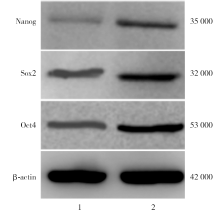
An experimental study on PD98059 reversing multiple drug resistance of human glioma stem cells by MEK/ERK signaling pathways
Wen Bobin, Gan Jie, Wang Zheng( )
)
Department of Neurosurgery ,Fourth Hospital of Changsha, Changsha Hospital of Integrated Traditinal Chinese and Western Medicine Changsha 410006, China
-
Received:2025-05-13Revised:2025-06-19Online:2025-12-08Published:2025-12-31 -
Contact:Wang Zheng E-mail:30694283@qq.com -
Supported by:Scientific Research Project of Health Commission in Hunan Province of China(202204045055)
Cite this article
Wen Bobin, Gan Jie, Wang Zheng. An experimental study on PD98059 reversing multiple drug resistance of human glioma stem cells by MEK/ERK signaling pathways[J]. Journal of International Oncology, 2025, 52(12): 737-744.
share this article
"
| 基因 | 引物 | 相对分子 质量 |
|---|---|---|
| MEK | 正向:5'-ACCAGGCAGAAATCAACGAC-3' | 224 000 |
| 反向:5'-GATGAACGTCCCAAAGCACT-3' | ||
| ERK | 正向:5'-TTACTGCGCTTCAGACATGAGA-3' | 111 000 |
| 反向:5'-ATCTGTTTCCATGAGGTCCTGT-3' | ||
| MDR1 | 正向:5'-CCCATCATTGCAATAGCAGG-3' | 167 000 |
| 反向:5'-GTTCAAACTTCTGCTCCTGA-3' | ||
| β-actin | 正向:5'-TCACCCACACTGTGCCCATCTACGA-3' | 295 000 |
| 反向:5'-CAGCGGAACCGCTCATTGCCAATGG-3' |
"

"
| 浓度(μmol/L) | 24 h | 48 h | 72 h | 96 h |
|---|---|---|---|---|
| 0 | 55.32±5.67 | 56.11±5.72 | 56.58±5.83 | 56.22±5.77 |
| 25 | 44.33±4.51a | 46.12±4.66a | 46.82±4.71a | 45.72±4.58a |
| 50 | 35.91±3.57ab | 42.55±4.38ab | 36.69±3.79ab | 42.51±4.37ab |
| 100 | 31.75±3.22abc | 39.11±4.09abc | 32.55±3.36abc | 40.01±4.06abc |
| 200 | 25.14±2.63abcd | 28.51±2.93abcd | 23.11±2.31abcd | 29.66±3.01abcd |
| 300 | 24.81±2.53abcd | 28.01±2.88abcd | 22.84±2.29abcd | 29.01±3.05abcd |
| 400 | 24.10±2.51abcd | 27.89±2.81abcd | 22.57±2.26abcd | 28.93±2.97abcd |
| F值 | 56.22 | 42.69 | 34.19 | 60.28 |
| P值 | <0.001 | <0.001 | <0.001 | <0.001 |
"
"
"
| 组别 | 多柔比星 | 长春新碱 |
|---|---|---|
| 对照组 | 18.21±0.33 | 22.07±1.51 |
| 抑制剂组 | 27.73±1.86a | 33.89±3.12a |
| 激活组 | 20.11±2.06ab | 25.41±2.65ab |
| MDR1敲除组 | 25.77±2.61ac | 30.19±3.08ac |
| F值 | 36.46 | 40.14 |
| P值 | <0.001 | <0.001 |
"
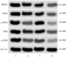
| 组别 | MEK | ERK | MDR1 |
|---|---|---|---|
| 对照组 | 1.00±0.00 | 1.00±0.00 | 1.00±0.00 |
| 抑制剂组 | 0.29±0.05a | 0.35±0.06a | 0.51±0.08a |
| 激活组 | 0.68±0.07ab | 0.74±0.07ab | 0.89±0.09ab |
| MDR1敲除组 | 0.33±0.03ac | 0.38±0.04ac | 0.56±0.06ac |
| F值 | 30.26 | 22.59 | 18.75 |
| P值 | <0.001 | <0.001 | <0.001 |
"

"
| 组别 | p-MEK/MEK | p-ERK/ERK | MDR1 |
|---|---|---|---|
| 对照组 | 0.90±0.09 | 1.19±0.13 | 1.08±0.12 |
| 抑制剂组 | 0.29±0.05a | 0.37±0.06a | 0.62±0.07a |
| 激活组 | 0.47±0.05ab | 0.55±0.06ab | 0.73±0.07ab |
| MDR1敲除组 | 0.32±0.04ac | 0.40±0.04ac | 0.65±0.06ac |
| F值 | 51.74 | 42.30 | 36.58 |
| P值 | <0.001 | <0.001 | <0.001 |
| [1] | Ramachandran R, Jeans AF. Breaking down glioma-microenvironment crosstalk[J]. Neuroscientist, 2025, 31(2): 177-194. DOI: 10.1177/10738584241259773. |
| [2] | Varela ML, Comba A, Faisal SM, et al. Gene therapy for high grade glioma: the clinical experience[J]. Expert Opin Biol Ther, 2023, 23(2): 145-161. DOI: 10.1080/14712598.2022.2157718. |
| [3] | Song KW, Lim M, Monje M. Complex neural-immune interactions shape glioma immunotherapy[J]. Immunity, 2025, 58(5): 1140-1160. DOI: 10.1016/j.immuni.2025.04.017. |
| [4] | Wu F, Yang J, Liu J, et al. Signaling pathways in cancer-associated fibroblasts and targeted therapy for cancer[J]. Signal Transduct Target Ther, 2021, 6(1): 218. DOI: 10.1038/s41392-021-00641-0. |
| [5] | Tsai TF, Lin JF, Lin YC, et al. Cisplatin contributes to programmed death-ligand 1 expression in bladder cancer through ERK1/2-AP-1 signaling pathway[J]. Biosci Rep, 2019, 39(9): BSR20190362. DOI: 10.1042/BSR20190362. |
| [6] |
Chen J, Liu G, Wang X, et al. Glioblastoma stem cell-specific histamine secretion drives pro-angiogenic tumor microenvironment remodeling[J]. Cell Stem Cell, 2022, 29(11): 1531-1546.e7. DOI: 10.1016/j.stem.2022.09.009.
pmid: 36265493 |
| [7] | Liu YP, Yang CJ, Huang MS, et al. Cisplatin selects for multidrug-resistant CD133+ cells in lung adenocarcinoma by activating notch signaling[J]. Cancer Res, 2013, 73(1): 406-416. DOI: 10.1158/0008-5472.CAN-12-1733. |
| [8] | 赵非, 黄松明, 丁桂霞, 等. 氧化应激介导的Ras-ERK信号通路活化参与醛固酮诱导的肾小球系膜细胞增殖[J]. 中华肾脏病杂志, 2012, 28(1): 41-46. DOI: 10.3760/cma.j.issn.1001-7097.2012.01.009. |
| [9] |
Miller JJ. Targeting IDH-mutant glioma[J]. Neurotherapeutics, 2022, 19(6): 1724-1732. DOI: 10.1007/s13311-022-01238-3.
pmid: 35476295 |
| [10] |
Sioutas G, Nikova A, Birbilis T. Risk factors for pediatric glioma[J]. Folia Med (Plovdiv), 2022, 64(4): 566-571. DOI: 10.3897/folmed.e64431.
pmid: 36045476 |
| [11] |
Karschnia P, Gerritsen J, Teske N, et al. The oncological role of resection in newly diagnosed diffuse adult-type glioma defined by the WHO 2021 classification: a review by the RANO resect group[J]. Lancet Oncol, 2024, 25(9): e404-e419. DOI: 10.1016/S1470-2045(24)00130-X.
pmid: 39214112 |
| [12] | Zhao L, Qiu Z, Yang Z, et al. Lymphatic endothelial-like cells promote glioblastoma stem cell growth through cytokine-driven cholesterol metabolism[J]. Nat Cancer, 2024, 5(1): 147-166. DOI: 10.1038/s43018-023-00658-0. |
| [13] | Hawly J, Murcar MG, Schcolnik-Cabrera A, et al. Glioblastoma stem cell metabolism and immunity[J]. Cancer Metastasis Rev, 2024, 43(3): 1015-1035. DOI: 10.1007/s10555-024-10183-w. |
| [14] |
Joyce T, Jagasia S, Tasci E, et al. An overview of CD133 as a functional unit of prognosis and treatment resistance in glioblastoma[J]. Curr Oncol, 2023, 30(9): 8278-8293. DOI: 10.3390/curroncol30090601.
pmid: 37754516 |
| [15] | 施佳, 董旭宸, 戴晓晓, 等. 胶质瘤初发与自然复发后肿瘤干细胞克隆及分子遗传学特征比较[J]. 中华神经医学杂志, 2019, 18(9): 865-874. DOI: 10.3760/cma.j.issn.1671-8925.2019.09.001. |
| [16] | 邓永文, 周向阳, 张明宇, 等. U251细胞系中脑肿瘤干细胞耐药性的初步研究[J]. 中国现代医学杂志, 2011, 21(7): 753-758, 765. DOI: 10.3969/j.issn.1005-8982.2011.07.001. |
| [17] |
Venere M, Fine HA, Dirks PB, et al. Cancer stem cells in gliomas: identifying and understanding the apex cell in cancer's hierarchy[J]. Glia, 2011, 59(8): 1148-1154. DOI: 10.1002/glia.21185.
pmid: 21547954 |
| [18] |
Hosseinali Z, Mohammadshahi J, Teimourpour A, et al. Molecular identification of multiple drug resistance (MDR) strain of mycobacterium tuberculosis[J]. Mol Biol Rep, 2023, 50(12): 10271-10275. DOI: 10.1007/s11033-023-08867-7.
pmid: 37971566 |
| [19] |
Kaushik M, Kaushik A, Jain A, et al. AmpC inhibition: an explicit approach against multi-drug resistance (MDR)[J]. Curr Top Med Chem, 2023, 23(20): 1919-1927. DOI: 10.2174/1568026623666230504095005.
pmid: 37150991 |
| [20] |
Koltai T. The complex relationship between multiple drug resistance and the tumor pH gradient: a review[J]. Cancer Drug Resist, 2022, 5(2): 277-303. DOI: 10.20517/cdr.2021.134.
pmid: 35800371 |
| [21] | 曾飞跃, 王延金, 陈风华, 等. MDR1在人脑肿瘤干细胞中的表达及意义[J]. 吉林大学学报(医学版), 2009, 35(5): 769-773. DOI: 10.13481/j.1671-587x.2009.05.062. |
| [22] | Wiwatchaitawee K, Mekkawy AI, Quarterman JC, et al. The MEK 1/2 inhibitor PD98059 exhibits synergistic anti-endometrial cancer activity with paclitaxel in vitro and enhanced tissue distribution in vivo when formulated into PAMAM-coated PLGA-PEG nanoparticles[J]. Drug Deliv Transl Res, 2022, 12(7): 1684-1696. DOI: 10.1007/s13346-021-01065-7. |
| [23] | 林芳, 马良娟. 金属蛋白酶在皮肤光老化信号通路中的研究进展[J]. 临床与病理杂志, 2018, 38(11): 2502-2507. DOI: 10.3978/j.issn.2095-6959.2018.11.033. |
| [1] | Hai Yanan, Bao Wenfang, Shentu Hangxiao, Chen Jingde. Mechanism of immunotherapy resistance and the progress of post-resistance treatment for dMMR/MSI-H metastatic colorectal cancer [J]. Journal of International Oncology, 2025, 52(9): 598-602. |
| [2] | Liao Zhipeng, He Yonglin, Du Aichao, Pan Yawen. Mechanisms of radionuclide therapy for tumors and research advances in gliomas [J]. Journal of International Oncology, 2025, 52(11): 720-725. |
| [3] | Dai Yujuan, Chen Xianying, Huang Wei, Chen Dachao. Analysis of influencing factors and construction of a risk prediction model for early death in adult glioma [J]. Journal of International Oncology, 2025, 52(10): 609-613. |
| [4] | Liu Pingping, He Xuefang, Zhang Yi, Yang Xu, Zhang Shanshan, Ji Yifei. Risk factors of postoperative recurrence in patients with primary brain glioma and prediction model construction [J]. Journal of International Oncology, 2024, 51(4): 193-197. |
| [5] | Yang Zhi, Lu Yiqiao, Gu Huayan, Ding Jialing, Guo Guilong. Research progress of tumor microenvironment mediated drug resistance in targeted therapy of breast cancer [J]. Journal of International Oncology, 2024, 51(4): 235-238. |
| [6] | Gong Yan, Chen Honglei. Research progress on the mechanism of microRNA regulation of cisplatin resistance in ovarian cancer [J]. Journal of International Oncology, 2024, 51(3): 186-190. |
| [7] | Du Aichao, Cheng Houxiang, Dai Junqiang, Pan Yawen. Advances in the study of the role of tumor treating fields therapy in the treatment of glioblastoma [J]. Journal of International Oncology, 2024, 51(10): 639-644. |
| [8] | An Rong, Liu Meihua, Wang Peichen, Wang Xiaohui. Research progress of Nrf2 in ovarian cancer [J]. Journal of International Oncology, 2023, 50(8): 493-497. |
| [9] | Liu Xiaojie, Huang Junxing. Research progress of NADPH oxidase 2 in malignant tumors [J]. Journal of International Oncology, 2023, 50(10): 618-621. |
| [10] | Wang Xi, Wu Chuanqing. Research progress in reversing multidrug resistance in colorectal cancer [J]. Journal of International Oncology, 2023, 50(1): 42-46. |
| [11] | Ma Xiaoping, Chang Junli, Sun Xingyuan, Yang Yanping. Study progression on mechanism of long non-coding RNAs regulating drug resistance in osteosarcoma [J]. Journal of International Oncology, 2023, 50(1): 51-54. |
| [12] | Xiao Nan, Sun Pengfei. Research progress of oxidative stress in the sensitivity of chemoradiotherapy for gliomas [J]. Journal of International Oncology, 2022, 49(6): 357-361. |
| [13] | Zhu Yishuo, Cui Yujie, Liu Qi, Li Jun, Fan Yuechao. Analysis of risk factors and prediction model establishment for early postoperative recurrence in glioma patients [J]. Journal of International Oncology, 2022, 49(2): 79-83. |
| [14] | Kong Chunyu, Sun Pengfei. SLC7A11 and glioma [J]. Journal of International Oncology, 2022, 49(10): 604-607. |
| [15] | Chen Peiyao, Jia Junmei. Mechanism and application of hypoxia affecting immunotherapy drug resistance [J]. Journal of International Oncology, 2021, 48(8): 489-493. |
| Viewed | ||||||
|
Full text |
|
|||||
|
Abstract |
|
|||||