Journal of International Oncology ›› 2026, Vol. 53 ›› Issue (3): 144-149.doi: 10.3760/cma.j.cn371439-20250415-00023
• Original Article • Previous Articles Next Articles
Evaluation of the risk of low-blood-flow BI-RADS category 4 breast lesions with an ultrasound-based XGBoost model
He Yuqing( ), Wu Zizheng, Qi Zhengqin
- Department of Ultrasound,First Hospital of Qinhuangdao,Qinhuangdao 066000,China
-
Received:2025-04-15Online:2026-03-08Published:2026-02-09 -
Contact:He Yuqing E-mail:347263253@qq.com -
Supported by:Hebei Provincial Medical Science Research Project(20231893);Qinhuangdao Science and Technology Research and Development Program(202301A199)
Cite this article
He Yuqing, Wu Zizheng, Qi Zhengqin. Evaluation of the risk of low-blood-flow BI-RADS category 4 breast lesions with an ultrasound-based XGBoost model[J]. Journal of International Oncology, 2026, 53(3): 144-149.
share this article
"
| 临床及超声 特征参数 | 良性患者 (n=174) | 恶性患者 (n=143) | χ²值 | P值 |
|---|---|---|---|---|
| 年龄(岁) | ||||
| <60 | 109(62.6) | 85(59.4) | 0.34 | 0.565 |
| ≥60 | 65(37.4) | 58(40.6) | ||
| 乳腺癌家族史 | ||||
| 无 | 161(92.5) | 128(89.5) | 0.89 | 0.346 |
| 有 | 13(7.5) | 15(10.5) | ||
| 肥胖 | ||||
| 是 | 137(78.7) | 102(71.3) | 2.32 | 0.128 |
| 否 | 37(21.3) | 41(28.7) | ||
| 饮酒史 | ||||
| 无 | 155(89.1) | 119(83.2) | 2.30 | 0.129 |
| 有 | 19(10.9) | 24(16.8) | ||
| 吸烟史 | ||||
| 无 | 162(93.1) | 129(90.2) | 0.87 | 0.350 |
| 有 | 12(6.9) | 14(9.8) | ||
| 血流分级 | ||||
| 低 | 101(58.0) | 65(45.5) | 4.99 | 0.026 |
| 高 | 73(42.0) | 78(54.5) | ||
| 病灶最大径(cm) | ||||
| ≤2 | 107(61.5) | 71(49.7) | 4.47 | 0.034 |
| >2 | 67(38.5) | 72(50.3) | ||
| 微钙化 | ||||
| 无 | 104(59.8) | 64(44.8) | 7.10 | 0.009 |
| 有 | 70(40.2) | 79(55.2) | ||
| 形态 | ||||
| 规则 | 76(43.7) | 48(33.6) | 3.37 | 0.066 |
| 不规则 | 98(56.3) | 95(66.4) | ||
| 边缘 | ||||
| 光整 | 74(42.5) | 57(39.9) | 0.23 | 0.631 |
| 不光整 | 100(57.5) | 86(60.1) | ||
| 内部回声 | ||||
| 均匀 | 82(47.1) | 51(35.7) | 4.24 | 0.041 |
| 不均匀 | 92(52.9) | 92(64.3) | ||
| 后方回声 | ||||
| 不衰减 | 106(60.9) | 49(34.3) | 22.32 | <0.001 |
| 衰减 | 68(39.1) | 94(65.7) | ||
| 平行位 | ||||
| 是 | 88(50.6) | 67(46.9) | 0.44 | 0.510 |
| 否 | 86(49.4) | 76(53.1) |
"
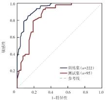
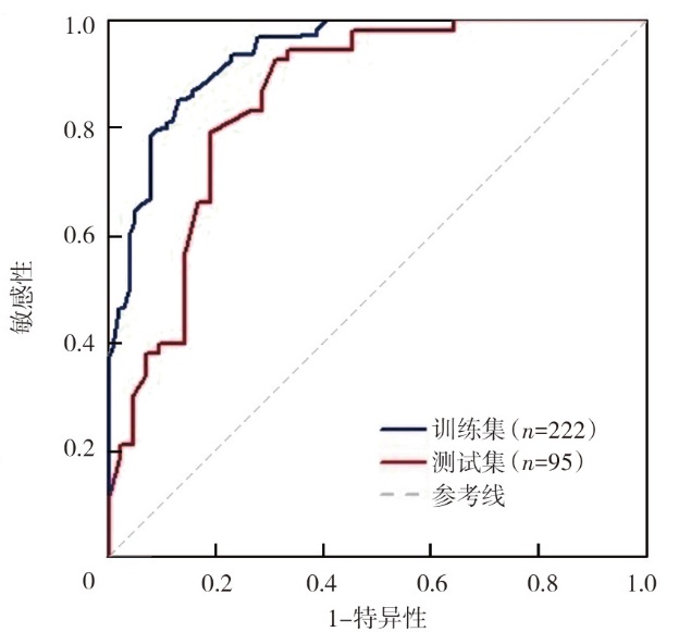
| 数据集 | AUC | 95%CI | 准确性(%) | 敏感性(%) | 特异性(%) | Kappa系数 |
|---|---|---|---|---|---|---|
| 训练集(n=222) | 0.936 | 0.902~0.965 | 86.0 | 88.5 | 83.2 | 0.715 |
| 测试集(n=95) | 0.852 | 0.787~0.906 | 76.8 | 78.6 | 75.0 | 0.533 |
"
"

"
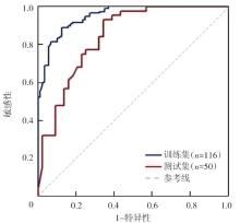
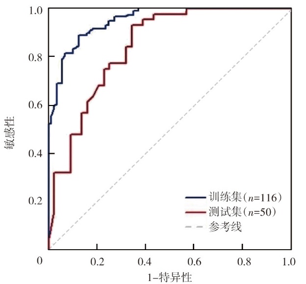
| 数据集 | AUC | 95%CI | 准确性(%) | 敏感性(%) | 特异性(%) | Kappa系数 |
|---|---|---|---|---|---|---|
| 训练集(n=116) | 0.951 | 0.917~0.975 | 86.5 | 87.9 | 84.8 | 0.717 |
| 测试集(n=50) | 0.843 | 0.766~0.904 | 79.6 | 81.5 | 77.8 | 0.591 |
"
"

"
| [1] | Sung H, Ferlay J, Siegel RL, et al. Global cancer statistics 2020: GLOBOCAN estimates of incidence and mortality worldwide for 36 cancers in 185 countries[J]. CA Cancer J Clin, 2021, 71(3): 209-249. DOI: 10.3322/caac.21660. |
| [2] | Avci T, Erkent M, Turnaolu H, et al. Are we on the side of over-diagnosis and treatment in BI-RADS 4A breast lesions?[J]. Ann Saudi Med, 2021, 28(3): 501-506. DOI: 10.5455/annalsmedres.2020.02.136. |
| [3] | Irmici G, Cozzi A, Depretto C, et al. Impact of an artificial intelligence decision support system among radiologists with different levels of experience in breast ultrasound: a prospective study in a tertiary center[J]. Eur J Radiol, 2025, 185: 112012. DOI: 10.1016/j.ejrad.2025.112012. |
| [4] | Zhou W, Luo H, Zhao H, et al. Unexpected breast cancer mimicking benign lesions on ultrasound-guided vacuum-assisted excision biopsy: a retrospective cross-sectional study over a 20-year period[J]. Front Oncol, 2023, 13: 1108689. DOI: 10.3389/fonc.2023.1108689. |
| [5] | Zhang X, Li H, Wang C, et al. Evaluating the accuracy of breast cancer and molecular subtype diagnosis by ultrasound image deep learning model[J]. Front Oncol, 2021, 11: 623506. DOI: 10.3389/fonc.2021.623506. |
| [6] | Zuopeng D, Weiyong L, Chunmei H, et al. Qualitative diagnosis of solid breast mass by blood flow in solid breast mass based on color doppler ultrasound[J]. J Med Imaging Health Inform, 2021, 11(6): 1608-1615. DOI: 10.1166/jmihi.2021.3682. |
| [7] | Gou F, Liu J, Xiao C, et al. Research on artificial-intelligence-assisted medicine: a survey on medical artificial intelligence[J]. Diagnostics (Basel), 2024, 14(14): 1472. DOI: 10.3390/diagnostics14141472. |
| [8] | Huang X, Cao J, Zu X. Tumor-associated macrophages: an important player in breast cancer progression[J]. Thorac Cancer, 2022, 13(3): 269-276. DOI: 10.1111/1759-7714.14268. |
| [9] |
Luo H, Li J, Shi Y, et al. Stiffness in breast masses with posterior acoustic shadowing: significance of ultrasound real time shear wave elastography[J]. BMC Med Imaging, 2022, 22(1): 71. DOI: 10.1186/s12880-022-00797-3.
pmid: 35430798 |
| [10] |
Kim S, Tran TXM, Song H, et al. Microcalcifications, mammographic breast density, and risk of breast cancer: a cohort study[J]. Breast Cancer Res, 2022, 24(1): 96. DOI: 10.1186/s13058-022-01594-0.
pmid: 36544167 |
| [11] | 何琴, 陈国珍, 武丽, 等. 乳腺肿块超声特征在乳腺癌筛查中的预测价值分析[J]. 中国妇幼卫生杂志, 2024, 15(6): 73-80. DOI: 10.19757/j.cnki.issn1674-7763.2024.06.011. |
| [12] | 杨海芳, 薛姣姣, 王鹏, 等. 彩色多普勒超声成像结合乳腺钼靶、EGF/EGFR、MIC-1对乳腺浸润性导管癌的评估价值[J]. 现代生物医学进展, 2024, 24(21): 4140-4143. DOI: 10.13241/j.cnki.pmb.2024.21.027. |
| [13] | 席芬, 张培培, 孝梦甦, 等. 乳腺错构瘤的临床与超声影像学特征分析[J]. 中华医学超声杂志(电子版), 2024, 21(5): 505-510. DOI: 10.3877/cma.j.issn.1672-6448.2024.05.009. |
| [14] |
Sha R, Kong XM, Li XY, et al. Global burden of breast cancer and attributable risk factors in 204 countries and territories, from 1990 to 2021: results from the Global Burden of Disease Study 2021[J]. Biomark Res, 2024, 12(1): 87. DOI: 10.1186/s40364-024-00631-8.
pmid: 39183342 |
| [1] | Chen Qiaoliang, Qin Xinyan, Lai Ruihe, Tan Shuangxiu. Diagnostic value of multimodal Nomogram model combining 18F-FDG PET/CT and ultrasound for triple negative breast cancer [J]. Journal of International Oncology, 2025, 52(9): 560-565. |
| [2] | Yu Cedric, Ren Lei, Lu Xiaoguang. Debates and reflection on modern proton radiotherapy and photon radiotherapy [J]. Journal of International Oncology, 2024, 51(7): 411-416. |
| [3] | Gu Fangmeng, Xu Chenyang, Lei Dapeng. Research progress on artificial intelligence-assisted electronic laryngoscopy in the diagnosis and treatment of laryngeal cancer and laryngeal precancerous lesions [J]. Journal of International Oncology, 2024, 51(5): 303-307. |
| [4] | Tan Shuangxiu, Zhang Yidan, Wang Ying, Yu Pengli, Kong Wentao, Yao Jing, Chen Qiaoliang. Value of conventional ultrasound combined with shear wave elastography in differentiating non-mass ductal carcinoma in situ from invasive breast cancer [J]. Journal of International Oncology, 2024, 51(12): 743-748. |
| [5] | Zhao Xin, Fan Xuewu, Tian Long, Hu Yimin. Application and evaluation study of 3D ultrasound in image guided radiotherapy for prostate cancer [J]. Journal of International Oncology, 2024, 51(1): 43-49. |
| [6] | Xie Yu, Jiang Cheng, Huang Mingmin, Guo Aibin, Yin Zhenyu, Lin Yongjuan. Effects of intrathecal infusion chemotherapy on intracranial pressure in non-small cell lung cancer patients with leptomeningeal metastases by ultrasound measurement of optic nerve sheath diameter [J]. Journal of International Oncology, 2023, 50(9): 532-539. |
| [7] | Feng Chengtian, Huang Furong, Cao Shiyu, Wang Jianyu, Nanding Abiyasi, Jiang Yongdong, Zhu Juanying. Relationships between HER2 protein expression and imaging features in HER2 positive breast cancer patients [J]. Journal of International Oncology, 2023, 50(9): 527-531. |
| [8] | Li Jiayi, Wang Yue, Shang Lanlan, Xu Xing, Zhao Yan. Practice and prospect of artificial intelligence in diagnosis and treatment of gastric cancer [J]. Journal of International Oncology, 2023, 50(11): 677-682. |
| [9] | Song Tongjun, Deng Rui, Fei Lei, Lei Jinhua, Cao Fengjun. Comparison of the efficacy and safety of percutaneous needle biopsy of pulmonary or pleural lesions guided by CT and ultrasound [J]. Journal of International Oncology, 2022, 49(9): 526-531. |
| [10] | Gao Junrong, Cao Manqing, Deng Yinghong. Advances in the diagnosis and treatment of hepatocellular carcinoma with contrast-enhanced ultrasound [J]. Journal of International Oncology, 2022, 49(7): 425-429. |
| [11] | Yan Danfang, Wang Lihong, Ye Hongxing, Yan Senxiang. Application of artificial intelligence in the target delineation of radiotherapy [J]. Journal of International Oncology, 2022, 49(3): 168-172. |
| [12] | Zhang Yumin, Zhao Xianwei, He Qianjin, Chen Jieneng. Value of contrast-enhanced ultrasound combined with serum CXCL8 and CXCR2 in the evaluation of postoperative efficacy of transcatheter arterial chemoembolization for primary liver cancer [J]. Journal of International Oncology, 2022, 49(10): 592-596. |
| [13] | Zheng Zhen, Zhu Zhaofeng. Application of endoscopic ultrasonography in the diagnosis of early esophageal cancer [J]. Journal of International Oncology, 2021, 48(3): 176-179. |
| Viewed | ||||||
|
Full text |
|
|||||
|
Abstract |
|
|||||