
Journal of International Oncology ›› 2025, Vol. 52 ›› Issue (11): 680-688.doi: 10.3760/cma.j.cn371439-20250513-00117
• Original article • Previous Articles Next Articles
Ma Lijun1,2, Ji Haitao1, Qu Xiaowei1, Wang Yanfeng1, Gao Xiaowei1( ), Han Jiming2
Received:2025-05-13
Revised:2025-10-01
Online:2025-11-08
Published:2025-12-21
Contact:
Gao Xiaowei
E-mail:43096886@qq.com
Supported by:Ma Lijun, Ji Haitao, Qu Xiaowei, Wang Yanfeng, Gao Xiaowei, Han Jiming. METTL3 inhibits the proliferation and migration of liver cancer cells by regulating the m6A modification of RAB5C[J]. Journal of International Oncology, 2025, 52(11): 680-688.
"
| 临床病理特征 | 例数 | RAB5C mRNA表达量 | χ2值 | P值 |
|---|---|---|---|---|
| 性别 | ||||
| 男 | 245 | 94.6(64.5,123.0) | 0.52 | 0.140 |
| 女 | 117 | 92.7(68.3,117.7) | ||
| 淋巴结转移状态 | ||||
| 未转移 | 252 | 93.9(65.1,120.4) | 0.72 | 0.020 |
| 转移 | 4 | 117.5(100.3,142.9) | ||
| TP53突变状态 | ||||
| 突变 | 105 | 100.2(80.5,133.1) | 0.74 | 0.010 |
| 未突变 | 105 | 84.7(62.3,115.9) |
"
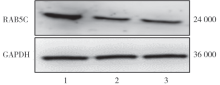
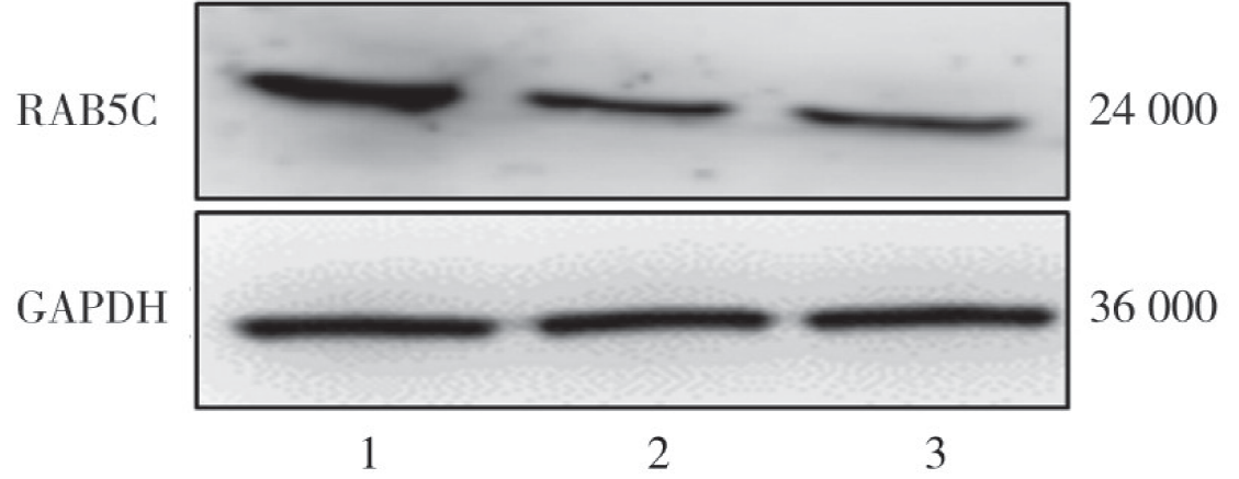
| 组别 | RAB5C mRNA | RAB5C蛋白 |
|---|---|---|
| si-NC组 | 1.00±0.12 | 1.00±0.01 |
| si-RAB5C-1组 | 0.18±0.05a | 0.36±0.11a |
| si-RAB5C-2组 | 0.12±0.06a | 0.40±0.07a |
| F值 | 108.02 | 59.02 |
| P值 | <0.001 | <0.001 |
"
"

"
"
"
"
"
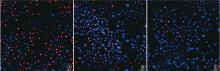
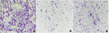
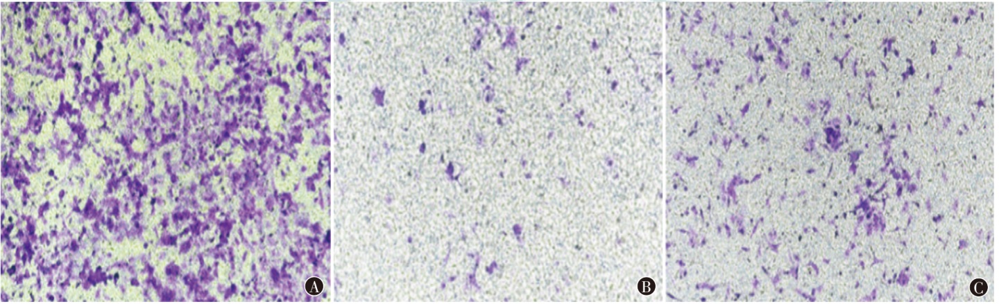
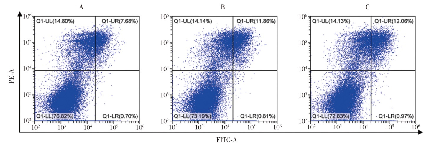
| 组别 | 细胞增殖率(%) | 细胞迁移率(%) | 细胞凋亡率(%) |
|---|---|---|---|
| si-NC+over-NC组 | 42.00±5.00 | 2.02±0.11 | 6.91±0.64 |
| si-METTL3+over-NC组 | 28.39±5.60a | 0.60±0.11a | 14.26±0.27a |
| si-METTL3+over-RAB5C组 | 40.00±4.06b | 1.98±0.09ab | 6.45±0.51ab |
| F值 | 6.72 | 170.70 | 194.40 |
| P值 | 0.030 | <0.001 | <0.001 |
"

"
"
| [1] | Zheng J, Wang S, Xia L, et al. Hepatocellular carcinoma: signaling pathways and therapeutic advances[J]. Signal Transduct Target Ther, 2025, 10(1): 35. DOI: 10.1038/s41392-024-02075-w |
| [2] | Su X, Yan X, Zhang H. The tumor microenvironment in hepatocellular carcinoma: mechanistic insights and therapeutic potential of traditional Chinese medicine[J]. Mol Cancer, 2025, 24(1):173. DOI: 10.1186/s12943-025-02378-8 |
| [3] | Zhang Z, Li J, He T, et al. The competitive endogenous RNA regulatory network reveals potential prognostic biomarkers for overall survival in hepatocellular carcinoma[J]. Cancer Sci, 2019, 110(9): 2905-2923. DOI: 10.1111/cas.14138. |
| [4] | Lai HH, Li CW, Hong CC, et al. TARBP2-mediated destabilization of Nanog overcomes sorafenib resistance in hepatocellular carcinoma[J]. Mol Oncol, 2019, 13(4): 928-945. DOI: 10.1002/1878-0261.12449. |
| [5] |
Jha A, van Zanten TS, Philippe JM, et al. Quantitative control of GPCR organization and signaling by endocytosis in epithelial morphogenesis[J]. Curr Biol, 2018, 28(10): 1570-1584.e6. DOI: 10.1016/j.cub.2018.03.068.
pmid: 29731302 |
| [6] | Parks XX, Ronzier E, O-Uchi J, et al. Fluvastatin inhibits Rab5-mediated IKs internalization caused by chronic Ca2+-dependent PKC activation[J]. J Mol Cell Cardiol, 2019, 129: 314-325. DOI: 10.1016/j.yjmcc.2019.03.016. |
| [7] |
Jin L, Huo Y, Zheng Z, et al. Down-regulation of Ras-related protein Rab 5C-dependent endocytosis and glycolysis in cisplatin-resistant ovarian cancer cell lines[J]. Mol Cell Proteomics, 2014, 13(11): 3138-3151. DOI: 10.1074/mcp.M113.033217.
pmid: 25096996 |
| [8] |
Onodera Y, Nam JM, Hashimoto A, et al. Rab5c promotes AMAP1-PRKD2 complex formation to enhance β1 integrin recycling in EGF-induced cancer invasion[J]. J Cell Biol, 2012, 197(7): 983-996. DOI: 10.1083/jcb.201201065.
pmid: 22734003 |
| [9] | Wang CJ, Xiao CW, You TG, et al. Interferon-α enhances antitumor activities of oncolytic adenovirus-mediated IL-24 expression in hepatocellular carcinoma[J]. Mol Cancer, 2012, 11: 31. DOI: 10.1186/1476-4598-11-31. |
| [10] | 何佩霈, 杨朝凤, 李杨. 肝癌肿瘤微环境与消融治疗的研究现状与展望[J]. 国际肿瘤学杂志, 2024, 51(10): 655-659. DOI: 10.3760/cma.j.cn371439-20240328-00110. |
| [11] | Parikh ND, Tayob N, Al-Jarrah T, et al. Barriers to surveillance for hepatocellular carcinoma in a multicenter cohort[J]. JAMA Netw Open, 2022, 5(7): e2223504. DOI: 10.1001/jamanetworkopen.2022.23504. |
| [12] |
Liu J, Wang X, Peng Z, et al. The effects of insulin pre-administration in mice exposed to ethanol: alleviating hepatic oxidative injury through anti-oxidative, anti-apoptotic activities and deteriorating hepatic steatosis through SRBEP-1c activation[J]. Int J Biol Sci, 2015, 11(5): 569-586. DOI: 10.7150/ijbs.11039.
pmid: 25892964 |
| [13] | Goubran M, Wang W, Indik S, et al. Isolation of a human betaretrovirus from patients with primary biliary cholangitis[J]. Viruses, 2022, 14(5): 886. DOI: 10.3390/v14050886. |
| [14] | Wang Z, Li F, Zhang H, et al. RAB5C, a new mRNA binding target of HuR, regulates breast cancer cell proliferation[J]. Cell Biol Int, 2023, 47(2): 374-382. DOI: 10.1002/cbin.11969. |
| [15] |
Zhang W, Ji W, Li T, et al. MiR-145 functions as a tumor suppressor in papillary thyroid cancer by inhibiting RAB5C[J]. Int J Med Sci, 2020, 17(13): 1992-2001. DOI: 10.7150/ijms.44723.
pmid: 32788878 |
| [16] | He PC, He C. m6A RNA methylation: from mechanisms to therapeutic potential[J]. EMBO J, 2021, 40(3): e105977. DOI: 10.15252/embj.2020105977. |
| [17] | Wu X, Zeng M, Wei Y, et al. METTL3 and METTL14-mediated N6-methyladenosine modification of SREBF2-AS1 facilitates hepatocellular carcinoma progression and sorafenib resistance through DNA demethylation of SREBF2[J]. Sci Rep, 2024, 14(1): 6155. DOI: 10.1038/s41598-024-55932-7. |
| [18] | Chen M, Wei L, Law CT, et al. RNA N6-methyladenosine methylt-ransferase-like 3 promotes liver cancer progression through YTHDF2-dependent posttranscriptional silencing of SOCS2[J]. Hepatology, 2018, 67(6): 2254-2270. DOI: 10.1002/hep.29683. |
| [1] | Huang Jinfa, Zheng Lianqiu, Wu Jinpiao, Liu Deting, Chen Huiling. Prediction model for post-TACE infection risk in elderly patients with liver cancer [J]. Journal of International Oncology, 2025, 52(8): 517-522. |
| [2] | Guo Junlong, Zou Ruiqi, Chen Shaoqiang, Liang Yuxin, Li Jing, Yong Sunan, He Yuting, Xie Xiaobing, Li Ping. Research progress of RNA m6A modification in breast cancer [J]. Journal of International Oncology, 2025, 52(8): 532-537. |
| [3] | Hao Chunhai. Efficacy of rapamycin arterial perfusion combined with 131I-FAP loaded dextran microspheres for interventional embolization in the treatment of rabbits with liver transplantation tumor [J]. Journal of International Oncology, 2025, 52(6): 353-359. |
| [4] | Yang Xiaobin, Jiang Jinquan. Predictive value of serum MMP-9, FABP5 combined with MRI for the efficacy and recurrence of primary liver cancer after percutaneous radiofrequency ablation [J]. Journal of International Oncology, 2025, 52(6): 360-365. |
| [5] | Xing Hui, Tan Ying, Wang Xiuzhen, Li Rui, Liu Xia. Predictive analysis of NLR and TNF-α level for the efficacy of TACE combined with microwave ablation therapy in patients with massive liver cancer [J]. Journal of International Oncology, 2025, 52(2): 101-106. |
| [6] | Chen Hongjian, Zhang Suqing. Study on the relationship between serum miR-24-3p, H2AFX and clinical pathological features and postoperative recurrence in liver cancer patients [J]. Journal of International Oncology, 2024, 51(6): 344-349. |
| [7] | Peng Qin, Cai Yuting, Wang Wei. Advances on KPNA2 in liver cancer [J]. Journal of International Oncology, 2024, 51(3): 181-185. |
| [8] | Sun Guobao, Yang Qian, Zhuang Qingchun, Gao Binbin, Sun Xiaogang, Song Wei, Sha Dan. Research progress on the histopathological growth patterns of colorectal liver metastasis [J]. Journal of International Oncology, 2024, 51(2): 114-118. |
| [9] | Jiang Qiong, Yu Jinhong. Research progress on the application of ultrasound-targeted microbubble destruction technology in the treatment of liver cancer [J]. Journal of International Oncology, 2024, 51(11): 723-727. |
| [10] | Zhang Dongqian, Zhang Haiguang, Zhang Xiaoru, Zheng Xuan, Han Sugui, Li Ying, Hao Chunhai. Application of miR-9 and miR-195-3p in the diagnosis of primary hepatic carcinoma and their changes before and after interventional therapy [J]. Journal of International Oncology, 2024, 51(10): 627-631. |
| [11] | Xiang Yuling, Tan Jiajie, Xiong Yuanguo, Zhao Lirong, Li Chen, Zhang Hong. Effects of Anhydroicaritin on the proliferation, migration and apoptosis of hepatocellular carcinoma cells [J]. Journal of International Oncology, 2023, 50(9): 513-519. |
| [12] | Li Jiaxuan, Feng Yinglu. Mechanism of action of glucocorticoid receptors in the growth of hepatoma cells [J]. Journal of International Oncology, 2023, 50(4): 241-243. |
| [13] | He Ting, Wang Xi, Zhang Huizhong, Liu Xinyang, Wang Huiping, Dong Ke. Diagnostic value of serum TIM-3 in patients with liver cancer [J]. Journal of International Oncology, 2022, 49(9): 537-542. |
| [14] | Hu Ru, Li Donglin, Yan Xuebing. Methyltransferase like protein 14 and tumor [J]. Journal of International Oncology, 2022, 49(8): 478-483. |
| [15] | Zhang Yumin, Zhao Xianwei, He Qianjin, Chen Jieneng. Value of contrast-enhanced ultrasound combined with serum CXCL8 and CXCR2 in the evaluation of postoperative efficacy of transcatheter arterial chemoembolization for primary liver cancer [J]. Journal of International Oncology, 2022, 49(10): 592-596. |
| Viewed | ||||||
|
Full text |
|
|||||
|
Abstract |
|
|||||