国际肿瘤学杂志 ›› 2025, Vol. 52 ›› Issue (11): 680-688.doi: 10.3760/cma.j.cn371439-20250513-00117

• 论著 • 上一篇    下一篇

METTL3通过调控RAB5C的m6A修饰抑制肝癌细胞增殖迁移

马利军1,2, 姬海涛1, 屈晓威1, 王延峰1, 高晓伟1(), 韩继明2   

  1. 1延安大学附属医院检验科,延安 716000
    2延安大学基础医学院,延安 716000
  • 收稿日期:2025-05-13 修回日期:2025-10-01 出版日期:2025-11-08 发布日期:2025-12-21
  • 通讯作者: 高晓伟 E-mail:43096886@qq.com
  • 基金资助:
    延安市科技计划项目(2023-SFGG-003);延安市科技计划项目(2022SLSFGG-022)

METTL3 inhibits the proliferation and migration of liver cancer cells by regulating the m6A modification of RAB5C

Ma Lijun1,2, Ji Haitao1, Qu Xiaowei1, Wang Yanfeng1, Gao Xiaowei1(), Han Jiming2   

  1. 1Department of Clinical Laboratory, Yan'an University Affiliated Hospital, Yan'an 716000, China
    2Yan'an University School of Basic Medicine, Yan'an 716000, China
  • Received:2025-05-13 Revised:2025-10-01 Online:2025-11-08 Published:2025-12-21
  • Contact: Gao Xiaowei E-mail:43096886@qq.com
  • Supported by:
    Science and Technology Plan Project of Yan'an City(2023-SFGG-003);Science and Technology Plan Project of Yan'an City(2022SLSFGG-022)

摘要:

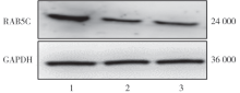
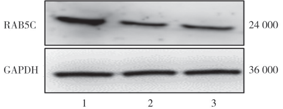
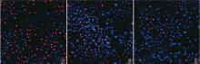
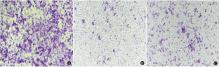
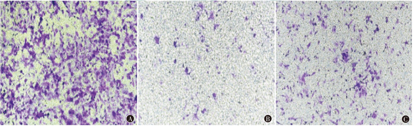
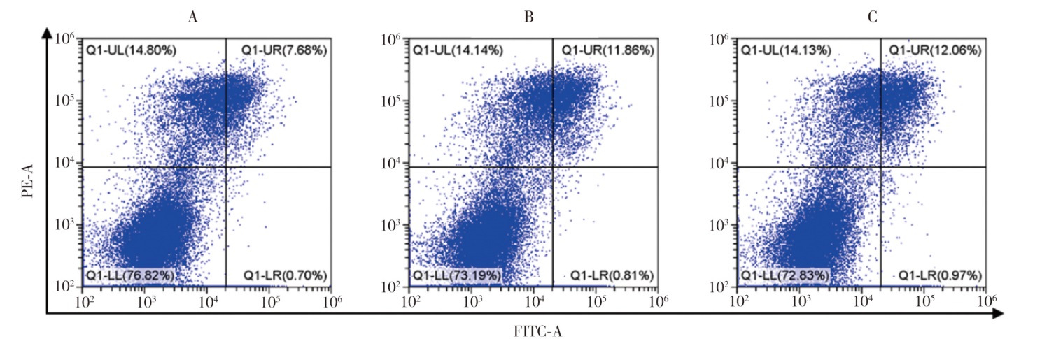
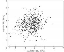
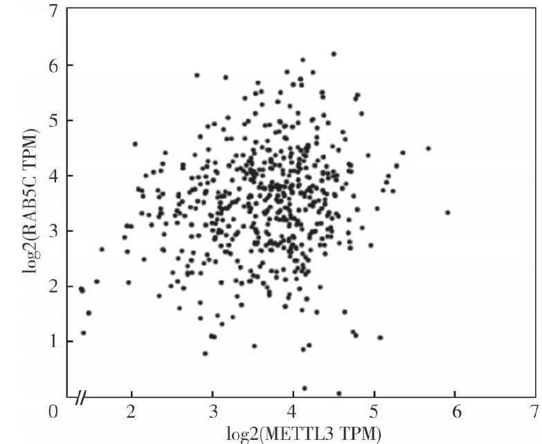
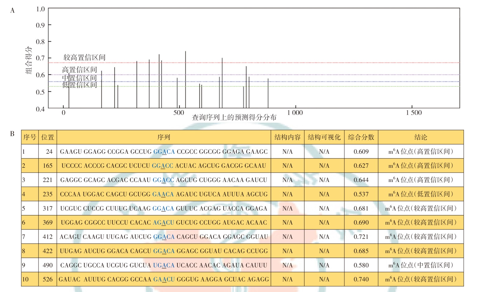
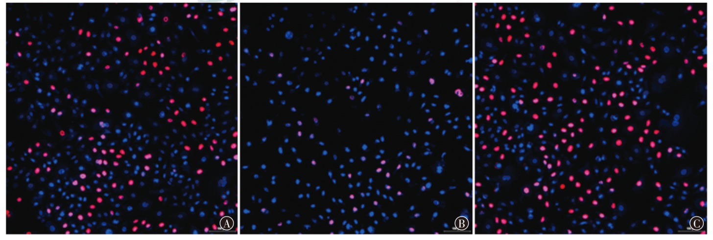
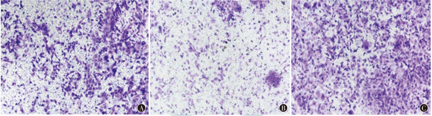
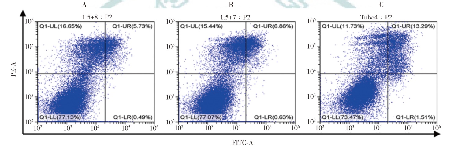
目的 探讨甲基转移酶样蛋白3(METTL3)通过调控RAB5C的稳定性对肝癌细胞增殖和迁移的影响及可能机制。方法 利用UALCAN数据库分析RAB5C在肝癌组织中的表达情况及其与临床病理特征的关系;将对数生长期的肝癌细胞株分为si-NC组(转染空白片段)、si-RAB5C-1组(沉默RAB5C)、si-RAB5C-2组(沉默RAB5C)、si-NC+over-NC组(转染空白片段+空白载体)、si-METTL3+over-NC组(沉默METTL3+空白载体)、si-METTL3+over-RAB5C组(沉默METTL3+RAB5C过表达载体)、RAB5C-WT+si-NC组(RAB5C野生型载体+空白载体)和RAB5C-MUT+si-METTL3组(RAB5C突变型载体+沉默METTL3);采用EdU实验检测细胞增殖能力;Transwell实验检测细胞迁移能力;流式细胞术检测细胞凋亡水平;使用GEPIA数据库分析METTL3与RAB5C表达的相关性;使用SARMP数据库对RAB5C中的m6A修饰位点进行预测;联合双荧光素酶报告基因实验与Me-RIP实验鉴定METTL3与RAB5C的靶向调控关系。结果 UALCAN数据库分析显示,RAB5C mRNA在正常组织(n=50)和肝癌组织(n=371)中的表达量分别为54.1(44.9,63.1)、93.5(67.1,120.0),差异有统计学意义(χ2=0.01,P<0.001)。RAB5C mRNA在淋巴结未转移(n=252)和转移(n=4)患者中的表达量分别为93.9(65.1,120.4)、117.5(100.3,142.9),在TP53突变(n=105)和未突变(n=105)患者中的表达量分别为100.2(80.5,133.1)、84.7(62.3,115.9),差异均有统计学意义(χ2=0.72,P=0.020;χ2=0.74,P=0.010)。EdU实验结果显示,si-NC组、si-RAB5C-1组和si-RAB5C-2组HepG2细胞增殖率分别为(39.3±2.93)%、(19.98±1.77)%、(19.98±2.46)%,差异有统计学意义(F=63.01,P<0.001),且si-RAB5C-1组和si-RAB5C-2组细胞增殖率均小于si-NC组(均P<0.05)。Transwell实验结果显示,si-NC组、si-RAB5C-1组、si-RAB5C-2组HepG2细胞迁移数分别为(94.2±1.2)、(26.1±0.5)、(25.1±0.6)个,差异有统计学意义(F=87.26,P<0.001),且si-NC组细胞迁移数均高于si-RAB5C-1组和si-RAB5C-2组(均P<0.05)。流式细胞术结果显示,si-NC组、si-RAB5C-1组和si-RAB5C-2组HepG2细胞凋亡率分别为(7.18±1.04)%、(12.56±1.50)%、(11.68±1.54)%,差异有统计学意义(F=13.09,P=0.007),且si-RAB5C-1组和si-RAB5C-2组细胞凋亡率均高于si-NC组(均P<0.05)。GEPIA、SARMP数据库分析显示,METTL3与RAB5C两者表达呈正相关(r=0.13,P=0.002),且RAB5C存在多个m6A修饰位点以及对应的m6A结合位点基序。MeRIP-qPCR实验结果显示,si-NC组HepG2细胞RAB5C相对表达量为1.00±0.11,高于si-METTL3组的0.28±0.18,差异有统计学意义(t=6.89,P=0.002)。双荧光素酶报告基因实验结果显示,与RAB5C-WT+si-NC组(1.00±0.16)相比,在肝癌HepG2细胞中沉默METTL3的表达可显著抑制野生型RAB5C启动子的荧光素酶活性(0.50±0.12),差异具有统计学意义(t=4.26,P=0.010);而与RAB5C-WT+si-NC组(1.00±0.03)相比,si-METTL3处理对突变型RAB5C启动子的荧光素酶活性(0.97±0.01)无显著影响(t=1.53,P=0.200)。EdU法、Transwell实验和流式细胞术结果显示,si-NC+over-NC组、si-METTL3+over-NC组和si-METTL3+over-RAB5C组细胞增殖、迁移和凋亡差异均具有统计学意义(均P<0.05);与si-METTL3+over-NC组比较,si-METTL3+over-RAB5C组细胞增殖、迁移和凋亡差异均有统计学意义(均P<0.05)。结论 METTL3可能通过介导m6A修饰调控RAB5C表达进而抑制肝癌的发生发展。

关键词: 肝肿瘤, 甲基转移酶样蛋白3, N6-甲基腺苷, RAB5C

Abstract:

Objective To explore the effects and possible mechanisms of methyltransferase-like protein 3 (METTL3) on the proliferation and migration of liver cancer cells by regulating the stability of RAB5C. Methods The expression of RAB5C in liver cancer tissues and its relationship with clinicopathological characteristics were were analyzed using the UALCAN database; The liver cancer cells of the logarithmic growth phase were divided into the following groups: si-NC group (transfected with a blank fragment), si-RAB5C-1 group (RAB5C silenced), si-RAB5C-2 group (RAB5C silenced), si-NC+over-NC group (transfected with a blank fragment+a blank vector), si-METTL3+over-NC group (METTL3 silenced+a blank vector), and si-METTL3+over-RAB5C group (METTL3 silenced+RAB5C overexpression vector); RAB5C-WT+si-NC group (RAB5C wild-type vector+control vector) and RAB5C-MUT+si-METTL3 group (RAB5C mutant vector+METTL3 silenced); EdU experiment was conducted to detect cell proliferation ability; Transwell experiment was conducted to detect cell migration ability; Flow cytometry was used to detect the level of cell apoptosis; GEPIA database was used to analyze the correlation between METTL3 and RAB5C expression; SARMP database was used to predict the m6A modification site in RAB5C; The targeted regulation relationship between METTL3 and RAB5C was identified by using dual luciferase reporter gene assay and Me-RIP assay. Results Analysis of the UALCAN database showed that the expression levels of RAB5C mRNA in normal tissues (n=50) and liver cancer tissues (n=371) were 54.1 (44.9, 63.1) and 93.5 (67.1, 120.0), respectively, with a statistically significant difference (χ2=0.01, P<0.001). The expression levels of RAB5C mRNA in patients with non-metastatic lymph nodes (n=252) and metastatic lymph nodes (n=4) were 93.9 (65.1, 120.4) and 117.5 (100.3, 142.9), respectively; the expression levels in patients with TP53 mutation (n=105) and non-mutation (n=105) were 100.2 (80.5, 133.1) and 84.7 (62.3, 115.9), respectively, with statistically significant differences (χ2=0.72, P=0.020; χ2=0.74, P=0.010). The EdU assay results indicated that the cell proliferation rates of the HepG2 cells in the si-NC group, si-RAB5C-1 group, and si-RAB5C-2 group were (39.3±2.93)%, (19.98±1.77)%, and (19.98±2.46)%, respectively, with a statistically significant difference (F=63.01, P<0.001), and the cell proliferation rates of the si-RAB5C-1 group and si-RAB5C-2 group were both lower than that of the si-NC group (both P<0.05). The results of the Transwell assay showed that the migration numbers of HepG2 cells in the si-NC group, si-RAB5C-1 group, and si-RAB5C-2 group were 94.2±1.2, 26.1±0.5, and 25.1±0.6, respectively, with a statistically significant difference (F=87.26, P<0.001), and the number of cell migration in the si-NC group was higher than that in the si-RAB5C-1 group and the si-RAB5C-2 group (both P<0.05). The results of flow cytometry showed that the apoptosis rates of HepG2 cells in the si-NC group, si-RAB5C-1 group and si-RAB5C-2 group were (7.18±1.04)%, (12.56±1.50)%, and (11.68±1.54)%, respectively, with a statistically significant difference (F=13.09, P=0.007), and the apoptosis rates of the si-RAB5C-1 group and the si-RAB5C-2 group were both higher than that of the si-NC group (both P<0.05). GEPIA and SARMP database analysis showed that the expression of METTL3 was positively correlated with that of RAB5C (r=0.13, P=0.002), and there were multiple m6A modification sites and corresponding m6A binding site motifs in RAB5C. The results of the MeRIP-qPCR experiment showed that the relative expression level of RAB5C in the HepG2 cell of si-NC group was 1.00±0.11, which was higher than that in the si-METTL3 group (0.28±0.18), with a statistically significant difference (t=6.89, P=0.002). The results of the dual-luciferase reporter gene assay showed that compared with the RAB5C-WT+si-NC group (1.00±0.16), silencing the expression of METTL3 in HepG2 liver cancer cells could significantly inhibit the luciferase activity of the wild-type RAB5C promoter (0.50±0.12), with a statistically significant difference (t=4.26, P=0.010); while compared with the RAB5C-WT+si-NC group (1.00±0.03), si-METTL3 treatment had no significant effect on the luciferase activity of the mutant RAB5C promoter (0.97±0.01) (t=1.53, P=0.200). The results of EdU assay, Transwell experiment, and flow cytometry showed that there were statistically significant differences in cell proliferation, migration, and apoptosis among the si-NC+over-NC group, si-METTL3+over-NC group, and si-METTL3+over-RAB5C group (all P<0.05). Compared with the si-METTL3+over-NC group, there were statistically significant differences in cell proliferation, migration, and apoptosis in the si-METTL3+over-RAB5C group (all P<0.05). Conclusions METTL3 may regulate the expression of RAB5C through mediated m6A modification, thus inhibiting the occurrence and development of liver cancer.

Key words: Liver neoplasms, Methyltransferase-like protein 3, N6-methyladenosine, RAB5C