国际肿瘤学杂志 ›› 2026, Vol. 53 ›› Issue (3): 144-149.doi: 10.3760/cma.j.cn371439-20250415-00023

• 论著 • 上一篇    下一篇

基于超声特征的XGBoost模型评估低血流分级BI-RADS 4类乳腺病变风险

贺玉卿(), 吴梓政, 齐铮琴   

  1. 秦皇岛市第一医院超声医学科,秦皇岛 066000
  • 收稿日期:2025-04-15 出版日期:2026-03-08 发布日期:2026-02-09
  • 通讯作者: 贺玉卿,Email: 347263253@qq.com
  • 基金资助:
    河北省医学科学研究课题计划(20231893);秦皇岛市科学技术研究与发展计划(202301A199)

Evaluation of the risk of low-blood-flow BI-RADS category 4 breast lesions with an ultrasound-based XGBoost model

He Yuqing(), Wu Zizheng, Qi Zhengqin   

  1. Department of Ultrasound,First Hospital of Qinhuangdao,Qinhuangdao 066000,China
  • Received:2025-04-15 Online:2026-03-08 Published:2026-02-09
  • Supported by:
    Hebei Provincial Medical Science Research Project(20231893);Qinhuangdao Science and Technology Research and Development Program(202301A199)

摘要:

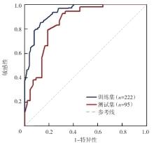
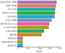
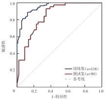
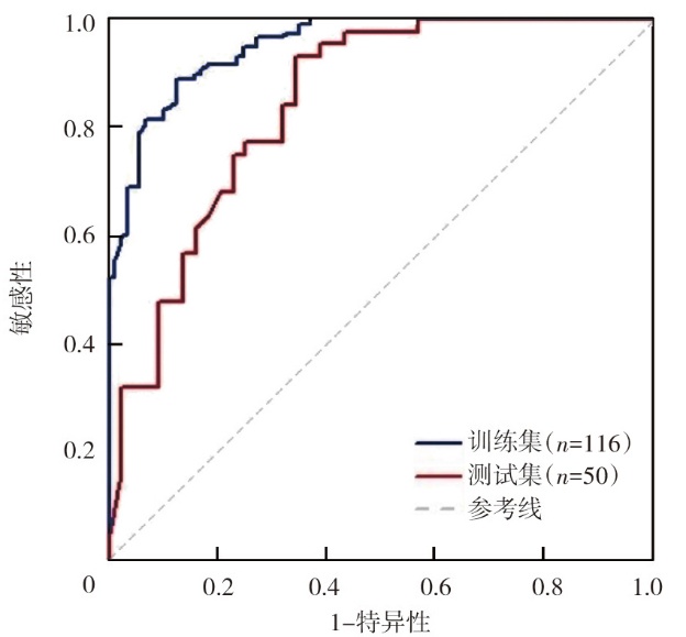
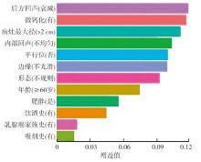
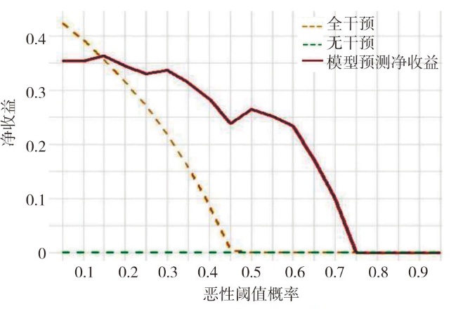
目的 基于临床和超声特征构建极端梯度提升(XGBoost)模型,评估模型预测低血流分级(Adler 0~Ⅰ级)乳腺影像报告与数据系统(BI-RADS)4类乳腺病变的恶性风险。方法 回顾性收集2023年6月至2024年12月于秦皇岛市第一医院诊断为BI-RADS 4类乳腺病变的317例女性患者的临床及超声资料(全样本),其中良性174例、恶性143例,采用7∶3随机分层抽样将患者分为训练集(n=222,良性122例、恶性100例)和测试集(n=95,良性52例、恶性43例);剔除高血流分级(Adler Ⅱ~Ⅲ级)患者后纳入低血流分级患者(低血流分级样本,n=166),同法按7∶3分为训练集(n=116,良性71例、恶性45例)和测试集(n=50,良性30例、恶性20例)。基于流行病学明确的乳腺癌风险因素(年龄、乳腺癌家族史、肥胖、饮酒史、吸烟史)及2013版ACR BI-RADS分类标准推荐的乳腺病变核心评估指标(血流分级、病灶最大径、微钙化、形态、边缘、内部回声、后方回声、平行位)构建预测BI-RADS 4类乳腺病变良、恶性的全样本XGBoost模型,剔除血流分级变量后余12项特征构建低血流分级XGBoost模型。采用受试者操作特征(ROC)曲线评估模型的预测效能,沙普利可加性解释(SHAP)分析明确模型特征贡献度,决策曲线分析(DCA)评估模型的准确性和实用性。结果 全样本中,良、恶性乳腺患者血流分级(χ²=4.99,P=0.026)、病灶最大径(χ²=4.47,P=0.034)、微钙化(χ²=7.10,P=0.009)、内部回声(χ²=4.24,P=0.041)、后方回声(χ²=22.32,P<0.001)分布差异均有统计学意义。ROC曲线分析显示,全样本训练集中XGBoost模型预测BI-RADS 4类乳腺病变良、恶性的曲线下面积(AUC)为0.936,95%CI为0.902~0.965,准确性为86.0%,敏感性为88.5%,特异性为83.2%;测试集AUC为0.852,95%CI为0.787~0.906,准确性为76.8%,敏感性为78.6%,特异性为75.0%。SHAP分析显示,血流分级(AdlerⅡ~Ⅲ级)对全样本XGBoost模型预测恶性风险的贡献最大,其次为边缘不光整、平行位否。低血流分级样本训练集中XGBoost模型预测BI-RADS 4类乳腺病变良、恶性的AUC为0.951,95%CI为0.917~0.975,准确性为86.5%,敏感性为87.9%,特异性为84.8%;测试集中AUC为0.843,95%CI为0.766~0.904,准确性为79.6%,敏感性为81.5%,特异性为77.8%。内部验证结果显示,XGBoost模型预测乳腺病变良、恶性的C-index为0.82。SHAP分析显示,后方回声衰减对低血流分级样本XGBoost模型预测恶性风险的正向贡献最大,其次为有微钙化、病灶最大径>2 cm、内部回声不均匀。DCA显示,该模型能提供较高的临床净获益,具有一定的临床实用性。结论 基于临床和超声特征的XGBoost模型可有效评估低血流分级BI-RADS 4类乳腺病变良、恶性,后方回声衰减、有微钙化、病灶最大径>2 cm、内部回声不均匀均为预测低血流分级BI-RADS 4类乳腺病变恶性风险的关键特征。

关键词: 乳腺疾病, 超声检查, 人工智能, XGBoost, BI-RADS

Abstract:

Objective To develop an extreme gradient boosting (XGBoost) model based on clinical and ultrasound features,and to evaluate the model's prediction of the malignancy risk of low-blood-flow (Adler grade 0 -Ⅰ) breast imaging-reporting and data system (BI-RADS) category 4 breast lesions. Methods Clinical and ultrasound data from 317 female patients diagnosed with BI-RADS category 4 breast lesions at First Hospital of Qinhuangdao from June 2023 to December 2024 were retrospectively collected (full-sample,174 benign,143 malignant). Patients were divided into a training set (n=222,122 benign,100 malignant) and a testing set (n=95,52 benign,43 malignant) using a 7∶3 stratified random sampling method. After excluding patients with high blood flow grades (Adler grade Ⅱ-Ⅲ),166 patients with low blood flow grades were collected and divided 7∶3 into training (n=116,71 benign,45 malignant) and testing (n=50,30 benign,20 malignant) sets. A full-sample XGBoost model for predicting the benign and malignant nature of BI-RADS category 4 breast lesions was constructed based on the well-defined epidemiological risk factors for breast cancer (age,family history of breast cancer,obesity,history of alcohol consumption,and smoking history) and the core assessment indicators for breast lesions recommended by the 2013 ACR BI-RADS classification standard (blood flow grade,maximum lesion diameter,microcalcification,shape,margin,internal echo,posterior echo,and parallel position). After excluding the blood flow grade variable,a low-blood-flow grade XGBoost model was constructed with the remaining 12 features. The predictive efficacy was evaluated using receiver operator characteristic (ROC) curves; SHapley additive explanation (SHAP) analysis was used to quantify feature contributions; decision curve analysis (DCA) was used to assess accuracy and practicability. Results There were statistically significant differences among patients with benign and malignant breast lesions in the full sample for blood flow grade (χ²=4.99,P=0.026),maximum lesion diameter (χ²=4.47,P=0.034),microcalcifications (χ²=7.10,P=0.009),internal echo (χ²=4.24,P=0.041),and posterior echo (χ²=22.32,P<0.001). ROC curve analysis showed that,for the full-sample training set,the area under the curve (AUC) of the XGBoost model for predicting benign and malignant BI-RADS category 4 breast lesions was 0.936 (95%CI: 0.902-0.965),with an accuracy of 86.0%,a sensitivity of 88.5%,and a specificity of 83.2%; for the testing set,the AUC was 0.852 (95%CI: 0.787-0.906),with an accuracy of 76.8%,a sensitivity of 78.6%,and a specificity of 75.0%. SHAP analysis showed that,the blood flow grade (Adler gradesⅡ-Ⅲ) had the greatest contribution to the prediction of malignancy risk by the XGBoost model for the full sample,followed by the irregularity of the margin and the absence of parallel position. For the low-blood-flow grade sample training set,the AUC of the XGBoost model for predicting benign and malignant BI-RADS category 4 breast lesions was 0.951 (95%CI: 0.917-0.975),with an accuracy of 86.5%,a sensitivity of 87.9%,and a specificity of 84.8%; for the testing set,the AUC was 0.843 (95%CI: 0.766-0.904),with an accuracy of 79.6%,a sensitivity of 81.5%,and a specificity of 77.8%. Internal validation results showed that the C-index of the XGBoost model for predicting benign and malignant breast lesions was 0.82. SHAP analysis showed that,the posterior echo attenuation had the greatest positive contribution to the prediction of malignancy risk by the XGBoost model for low-blood-flow grade samples,followed by the presence of microcalcification,maximum lesion diameter >2 cm,and inhomogeneous internal echo. DCA showed that this prediction model could provide high clinical net benefit and had certain clinical practicability. Conclusions The XGBoost model based on clinical and ultrasound features effectively evaluates benign and malignant nature of low-blood-flow BI-RADS category 4 breast lesions. Posterior echo attenuation,microcalcification,maximum lesion diameter >2 cm,and inhomogeneous internal echo are key features for predicting malignancy risk in low-blood-flow BI-RADS category 4 breast lesions.

Key words: Breast diseases, Ultrasonography, Artificial intelligence, XGBoost, BI-RADS