Journal of International Oncology ›› 2026, Vol. 53 ›› Issue (9): 540-546.doi: 10.3760/cma.j.cn371439-20260118-00086
• Original Article • Previous Articles Next Articles
Application value of multimodal ultrasound imaging in the diagnosis of benign and malignant cervical lesions
Shen Xuejie( ), Niu Caihong, Xu Ping, Xie Nan, Liu Yiran
Department of Ultrasound ,First Hospital of Qinhuangdao Qinhuangdao 066000, China
-
Received:2026-01-18Online:2026-09-08Published:2026-08-25 -
Contact:Shen Xuejie, Niu Caihong, Xu Ping, Xie Nan, Liu Yiran E-mail:shenxuejie123456@126.com -
Supported by:Science and Technology Program of Qinhuangdao of China(202501A171)

Cite this article
Shen Xuejie, Niu Caihong, Xu Ping, Xie Nan, Liu Yiran. Application value of multimodal ultrasound imaging in the diagnosis of benign and malignant cervical lesions[J]. Journal of International Oncology, 2026, 53(9): 540-546.
share this article
"
"

"
| 检查方法 | 病理结果 | 合计 | |
|---|---|---|---|
| 恶性 | 良性 | ||
| 常规二维超声 | |||
| 恶性 | 21 | 20 | 41 |
| 良性 | 10 | 43 | 53 |
| TVCDS | |||
| 恶性 | 23 | 12 | 35 |
| 良性 | 8 | 51 | 59 |
| 3D-PDUS | |||
| 恶性 | 25 | 8 | 33 |
| 良性 | 6 | 55 | 61 |
| SWE | |||
| 恶性 | 27 | 7 | 34 |
| 良性 | 4 | 56 | 60 |
| 多模态超声成像 | |||
| 恶性 | 31 | 1 | 32 |
| 良性 | 0 | 62 | 62 |
"
| 检查方法 | Kappa值 | 敏感性 [%(例数/总例数)] | 特异性 [%(例数/总例数)] | 准确性 [%(例数/总例数)] | 阳性预测值 [%(例数/总例数)] | 阴性预测值 [%(例数/总例数)] |
|---|---|---|---|---|---|---|
| 常规二维超声 | 0.333 | 67.74(21/31) | 68.25(43/63) | 68.09(64/94) | 51.22(21/41) | 81.13(43/53) |
| TVCDS | 0.534 | 74.19(23/31) | 80.95(51/63) | 78.72(74/94) | 65.71(23/35) | 86.44(51/59) |
| 3D-PDUS | 0.669 | 80.65(25/31) | 87.30(55/63)a | 85.11(80/94)a | 75.76(25/33)a | 90.16(55/61) |
| SWE | 0.742 | 87.10(27/31) | 88.89(56/63)a | 88.30(83/94)a | 79.41(27/34)a | 93.33(56/60)a |
| 多模态超声成像 | 0.976 | 100(31/31)abcd | 98.41(62/63)abcd | 98.94(93/94)abcd | 96.88(31/32)abcd | 100(62/62)abcd |
"
| 超声参数 | 宫颈炎(n=38) | 宫颈上皮内瘤变(n=25) | 宫颈癌(n=31) | F/U值 | P值 |
|---|---|---|---|---|---|
| TVCDS($\bar{x}±s$) | |||||
| PSV(cm/s) | 10.35±2.21 | 13.84±2.61a | 17.29±3.07ab | 59.86 | <0.001 |
| EDV(cm/s) | 5.56±1.03 | 5.91±1.22 | 6.05±1.36 | 1.54 | 0.220 |
| RI | 0.72±0.20 | 0.63±0.17a | 0.52±0.12ab | 11.93 | <0.001 |
| 3D-PDUS[M(Q1,Q3)] | |||||
| FI | 44.48(37.79,49.45) | 54.05(40.13,65.40)a | 57.78(45.54,71.03)a | 16.60 | <0.001 |
| VI | 1.20(0.87,1.89) | 3.27(1.54,4.24)a | 5.23(2.98,14.77)ab | 55.97 | <0.001 |
| VFI | 0.57(0.25,0.96) | 2.04(0.74,2.85)a | 9.31(5.25,19.82)ab | 52.18 | <0.001 |
| SWE(kPa,$\bar{x}±s$) | |||||
| Emean | 20.81±5.61 | 30.05±9.06a | 80.39±9.27ab | 527.62 | <0.001 |
| Emax | 27.16±7.14 | 35.53±8.53a | 90.13±8.88ab | 568.76 | <0.001 |
"
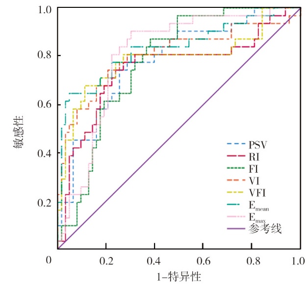
| 指标 | 截断值 | 敏感性(%) | 特异性(%) | AUC | 95%CI | P值 |
|---|---|---|---|---|---|---|
| PSV | 14.57 cm/s | 77.42 | 71.43 | 0.78 | 0.68~0.88 | <0.001 |
| RI | 0.62 | 77.42 | 73.02 | 0.75 | 0.63~0.87 | <0.001 |
| FI | 48.84 | 87.10 | 61.90 | 0.78 | 0.68~0.87 | <0.001 |
| VI | 4.74 | 74.19 | 79.37 | 0.81 | 0.70~0.91 | <0.001 |
| VFI | 3.74 | 67.74 | 88.89 | 0.79 | 0.68~0.90 | <0.001 |
| Emean | 57.70 kPa | 64.52 | 95.24 | 0.83 | 0.74~0.93 | <0.001 |
| Emax | 45.11 kPa | 90.32 | 69.84 | 0.81 | 0.71~0.90 | <0.001 |
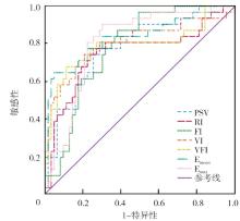
"
| [1] | 李锦鑫, 顾芬芬. 信迪利单抗联合多西他赛治疗宫颈癌疗效及对实验室指标的影响[J]. 国际肿瘤学杂志, 2025, 52(6): 366-373. DOI: 10.3760/cma.j.cn371439-20250225-00062. |
| [2] | Sahasrabuddhe VV. Cervical cancer: precursors and prevention[J]. Hematol Oncol Clin North Am, 2024, 38(4): 771-781. DOI: 10.1016/j.hoc.2024.03.005. |
| [3] | Zhu Y, Tang Y, Jiang Z, et al. Potential diagnostic value of quantitative superb microvascular imaging in premalignant and malignant cervical lesions[J]. Front Oncol, 2023, 13: 1250842. DOI: 10.3389/fonc.2023.1250842. |
| [4] | Tian Y, Luo H. Diagnostic accuracy of transvaginal ultrasound examination for local staging of cervical cancer: a systematic review and meta-analysis[J]. Med Ultrason, 2022, 24(3): 348-355. DOI: 10.11152/mu-3246. |
| [5] | 吴青京, 陈昌钊, 李波, 等. 经阴道超声和三维彩色血管能量成像单用及联用对宫颈癌分期的诊断效能研究[J]. 中国超声医学杂志, 2022, 38(1): 87-91. DOI: 10.3969/j.issn.1002-0101.2022.01.028. |
| [6] | Sainz JA, Castro L, Romo JM, et al. Evaluation of pre-malignant lesions of the uterine cervix by shear wave elastography: a new diagnostic tool[J]. Ultrasound Med Biol, 2021, 47(11): 3275-3282. DOI: 10.1016/j.ultrasmedbio.2021.06.021. |
| [7] | 谢幸, 孔北华, 段涛. 妇产科学[M]. 9版. 北京: 人民卫生出版社, 2018: 247-248, 295. |
| [8] | 中国抗癌协会妇科肿瘤专业委员会. 子宫颈癌诊断与治疗指南(2021年版)[J]. 中国癌症杂志, 2021, 31(6): 474-489. DOI: 10.19401/j.cnki.1007-3639.2021.06.06. |
| [9] | 中国医师协会超声医师分会. 中国妇科超声检查指南[M]. 北京: 人民卫生出版社, 2017. |
| [10] | Wang W, Gao X, Zhu Y, et al. Diagnostic significance of a color doppler ultrasound combined with serum CXCL16 and E-cad in cervical cancer[J]. Transl Cancer Res, 2021, 10(3): 1492-1499. DOI: 10.21037/tcr-21-123. |
| [11] | Guo YJ, Yan XJ, Zhao HX, et al. Quantitative evaluation of the normal cervix, cervical cancer, and cervical precancerous changes via real-time shear wave elastography[J]. J Ultrasound Med, 2023, 42(2): 345-354. DOI: 10.1002/jum.15981. |
| [12] | 温道浩, 赵鹏, 王乐, 等. 基于常规超声和三维能量多普勒的列线图预测宫颈癌前病变的临床价值[J]. 临床超声医学杂志, 2025, 27(3): 215-220. |
| [13] | Yang Y, Xu L, Yuan S, et al. Optimal screening and detection strategies for cervical lesions: a retrospective study[J]. J Cancer, 2024, 15(11): 3612-3624. DOI: 10.7150/jca.96128. |
| [14] | Li H, Li L, Sun J, et al. Value of TCT combined with serum CA153 and CA50 in early diagnosis of cervical cancer and precancerous lesions[J]. Pak J Med Sci, 2022, 38(6): 1471-1476. DOI: 10.12669/pjms.38.6.5503. |
| [15] | 朱一平, 郭道宁, 王亮, 等. 腹部超声、阴道超声联合剪切波弹性成像诊断宫颈癌的价值[J]. 中国临床医学影像杂志, 2021, 32(7): 517-520. DOI: 10.12117/jccmi.2021.07.014. |
| [16] | 张文莉, 冯彩霞, 王彩丽, 等. 3D-CPA对宫颈癌的检测结果及与miR-455、VEGF、MVD表达的相关性研究[J]. 中国医药导刊, 2022, 24(12): 1217-1221. DOI: 10.3969/j.issn.1009-0959.2022.12.013. |
| [17] | Zhang L, Li J, Shang Y, et al. The diagnostic value of transvaginal sonoelastography for cervical cancer and cervical intraepithelial neoplasia: a systematic review and meta-analysis[J]. Eur J Med Res, 2025, 30(1): 516. DOI: 10.1186/s40001-025-02777-z. |
| [18] | Liu B, Wei L, Wu L, et al. Predictive value of Netrin-1 expression and ultrasonic blood flow in cervical intraepithelial neoplasia severity[J]. Int J Womens Health, 2025, 17: 43-51. DOI: 10.2147/IJWH.S492376. |
| [1] | Zhang Xuhui, Liang Zhimin, Jiang Jing, Zhang Yan, Feng Jingbo. Relationship between CD4+ T cell TIM-3,PD-1 levels and the efficacy of cervical cancer immunotherapy,and the mediating role of macrophage polarization [J]. Journal of International Oncology, 2026, 53(9): 527-539. |
| [2] | Han Yufei, Li Fengmei, Zhang Weina, Zhang Ping, Hou Ping. Short-term efficacy and clinical outcomes of concurrent chemoradiotherapy followed by anlotinib maintenance treatment in patients with locally advanced cervical cancer [J]. Journal of International Oncology, 2026, 53(4): 219-223. |
| [3] | Yang Qing, He Xiyan, Sun Xiaotong. Research progress of ferroptosis in cervical cancer [J]. Journal of International Oncology, 2026, 53(3): 187-192. |
| [4] | Li Jinxin, Gu Fenfen. Efficacy of sintilimab combined with docetaxel in the treatment of cervical cancer and its impact on laboratory indicators [J]. Journal of International Oncology, 2025, 52(6): 366-373. |
| [5] | Zhou Xiaoyu, Pu Xuefeng, Long Shulin, Li Lu, He Wenying. Changes of T lymphocyte subsets and the relationship with postoperative lymph node metastasis in patients with stage Ⅰ and Ⅱ cervical cancer [J]. Journal of International Oncology, 2025, 52(4): 224-230. |
| [6] | Han Tao, Jia Peipei, Lu Jing. Predictive value of iRhom1,iRhom2 and TNF-α levels for the prognosis of patients with cervical cancer [J]. Journal of International Oncology, 2025, 52(3): 158-162. |
| [7] | Zumurelaiti·Ainiwa'er , Xian Jun, Han Lili. Diagnostic value of MRI combined with serum AIF-1 and NDRG4 for cervical cancer [J]. Journal of International Oncology, 2025, 52(12): 777-781. |
| [8] | Hou Xiaoxuan, Tang Wenjie, Xie Peng. Mechanism and research progress of ferroptosis in cervical cancer treatment [J]. Journal of International Oncology, 2025, 52(12): 791-794. |
| [9] | Ma Yongjia, Peng Siyu, Sun Pengfei. Research progress on combined radiotherapy, chemotherapy, and immunotherapy for cervical cancer [J]. Journal of International Oncology, 2025, 52(11): 726-731. |
| [10] | Wang Yan. Differential diagnostic value of transvaginal ultrasound elastography combined with serum miR-144-3p,CMTM6 for cervical precancerous lesions and cervical cancer [J]. Journal of International Oncology, 2024, 51(8): 504-509. |
| [11] | Peng Dan, Lyu Lu, Sun Pengfei. Research progress of radiomics in cervical cancer [J]. Journal of International Oncology, 2024, 51(8): 532-537. |
| [12] | Zhang Ningning, Yang Zhe, Tan Limei, Li Zhenning, Wang Di, Wei Yongzhi. Diagnostic value of cervical cell DNA ploidy analysis combined with B7-H4 and PKCδ for cervical cancer [J]. Journal of International Oncology, 2024, 51(5): 286-291. |
| [13] | Tan Shuangxiu, Zhang Yidan, Wang Ying, Yu Pengli, Kong Wentao, Yao Jing, Chen Qiaoliang. Value of conventional ultrasound combined with shear wave elastography in differentiating non-mass ductal carcinoma in situ from invasive breast cancer [J]. Journal of International Oncology, 2024, 51(12): 743-748. |
| [14] | Zhang Lu, Jiang Hua, Lin Zhou, Ma Chenying, Xu Xiaoting, Wang Lili, Zhou Juying. Analysis of curative effect and prognosis of immune checkpoint inhibitor in the treatment of recurrent and metastatic cervical cancer [J]. Journal of International Oncology, 2023, 50(8): 475-483. |
| [15] | Lyu Lu, Sun Pengfei. Gut flora and cervical cancer [J]. Journal of International Oncology, 2023, 50(6): 373-376. |
| Viewed | ||||||
|
Full text |
|
|||||
|
Abstract |
|
|||||