Journal of International Oncology ›› 2026, Vol. 53 ›› Issue (6): 321-330.doi: 10.3760/cma.j.cn371439-20251128-00053
• Original Article • Previous Articles Next Articles
Role and molecular mechanism of the Wnt/β-catenin signaling pathway in the regulation of NSCLC A549 cell proliferation and apoptosis by oleanolic acid
Wang Junqi1,2, Li Kairui1, Yuan Rong1,2, Lu Ying1,2, Xu Zhaojun2,3, Song Lan1,2( )
)
- 1
Medicine School ,Hunan University of Chinese Medicine Changsha 410208, China
2Hunan Provincial Key Laboratory of Vascular Biology and Translational Medicine ,Hunan University of Chinese Medicine Changsha 410208, China
3Department of Cardiothoracic Surgery ,First Hospital of Hunan University of Chinese Medicine Changsha 410007, China
-
Received:2025-11-28Online:2026-06-08Published:2026-06-05 -
Contact:Song Lan E-mail:songlan311492@hnucm.edu.cn -
Supported by:Natural Science Foundation of Hunan Province of China(2025JJ80944);Science Research Project of the Education Department of Hunan Province of China(22A0243);Innovation Project for Postgraduates of Hunan Province of China(LXBZZ2024168)
Cite this article
Wang Junqi, Li Kairui, Yuan Rong, Lu Ying, Xu Zhaojun, Song Lan. Role and molecular mechanism of the Wnt/β-catenin signaling pathway in the regulation of NSCLC A549 cell proliferation and apoptosis by oleanolic acid[J]. Journal of International Oncology, 2026, 53(6): 321-330.
share this article
"
"
"
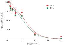
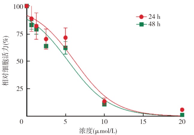
| 浓度(μmol/L) | 24 h | 48 h |
|---|---|---|
| 0(阴性对照) | 100.00±0.00 | 100.00±0.00 |
| 0.625 | 88.63±11.39a | 82.80±3.87ac |
| 1.25 | 81.91±11.92ab | 78.92±4.47abc |
| 2.5 | 70.14±9.19ab | 63.68±1.65abc |
| 5 | 71.37±8.78a | 62.10±5.81ab |
| 10 | 13.60±3.12ab | 11.03±1.32abc |
| 20 | 6.05±0.16ab | 1.16±0.38abc |
| F值 | 107.57 | 656.96 |
| P值 | <0.001 | <0.001 |
"

"
"
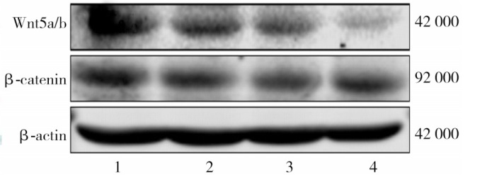
| 组别 | Wnt5a/b | β-catenin |
|---|---|---|
| 0 μmol/L OA | 0.99±0.02 | 0.99±0.02 |
| 1.25 μmol/L OA | 0.78±0.03a | 0.80±0.07a |
| 5 μmol/L OA | 0.67±0.03ab | 0.65±0.03ab |
| 0.004 g/L顺铂 | 0.68±0.03ac | 0.82±0.05ac |
| F值 | 80.55 | 26.37 |
| P值 | <0.001 | <0.001 |
"

"
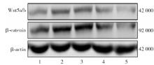
| 组别 | Wnt5a/b | β-catenin |
|---|---|---|
| 阴性对照组 | 1.03±0.06 | 0.99±0.02 |
| HLY78组 | 1.42±0.02a | 1.22±0.08a |
| OA组 | 0.98±0.07ab | 0.69±0.16ab |
| HLY78+OA组 | 1.17±0.05bc | 0.85±0.12abc |
| IWR-1组 | 0.34±0.10abcd | 0.39±0.19abcd |
| F值 | 116.70 | 19.20 |
| P值 | <0.001 | <0.001 |
"
"
"
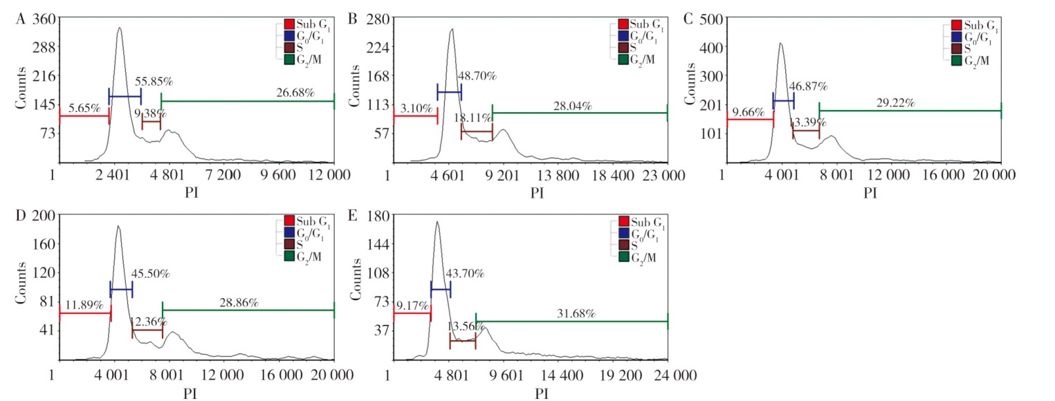
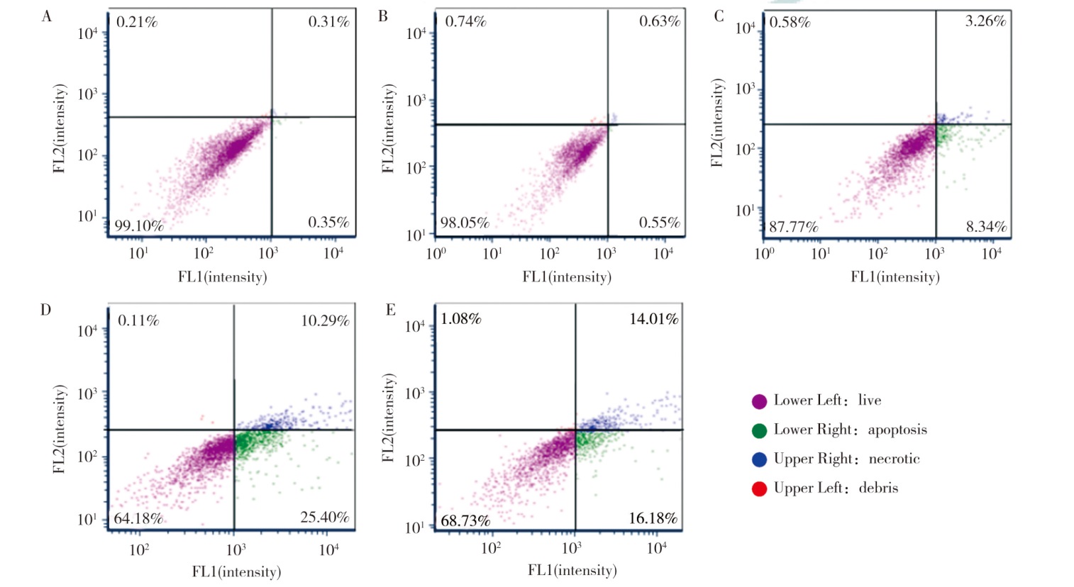
| 组别 | Sub G1期 | G0/G1期 | S期 | G2/M期 |
|---|---|---|---|---|
| 阴性对照组 | 5.45±0.21 | 55.46±0.37 | 10.81±1.24 | 26.64±0.15 |
| HLY78组 | 3.30±0.20a | 45.29±3.34a | 18.53±0.92a | 30.38±2.38 |
| OA组 | 9.69±1.91ab | 45.07±0.42ab | 13.80±2.09ab | 27.89±0.87 |
| HLY78+OA组 | 8.29±1.16abc | 46.15±2.61abc | 15.17±1.53abc | 28.37±3.73 |
| IWR-1组 | 8.21±1.26d | 52.15±4.93d | 11.73±2.76d | 25.94±2.85 |
| F值 | 15.16 | 7.86 | 8.36 | 1.56 |
| P值 | <0.001 | 0.004 | 0.003 | 0.260 |
"
"
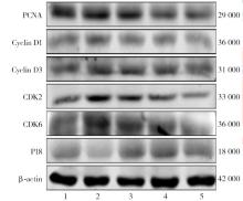
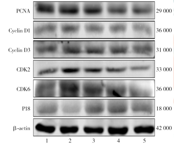
| 组别 | PCNA | Cyclin D1 | Cyclin D3 | CDK2 | CDK6 | P18 |
|---|---|---|---|---|---|---|
| 阴性对照组 | 0.99±0.01 | 0.98±0.02 | 0.98±0.01 | 0.99±0.02 | 0.99±0.02 | 0.99±0.01 |
| HLY78组 | 1.30±0.14a | 1.31±0.18a | 1.15±0.02a | 1.28±0.13a | 1.52±0.13a | 0.80±0.12a |
| OA组 | 0.81±0.07ab | 0.72±0.08ab | 0.81±0.02ab | 0.74±0.09ab | 0.43±0.09ab | 1.42±0.14ab |
| HLY78+OA组 | 1.02±0.09abc | 0.78±0.14abc | 0.93±0.05abc | 0.95±0.21abc | 1.00±0.03abc | 1.17±0.10abc |
| IWR-1组 | 0.75±0.04d | 0.61±0.09d | 0.83±0.03d | 0.38±0.03d | 0.33±0.04d | 1.26±0.06d |
| F值 | 20.23 | 17.19 | 63.77 | 24.62 | 127.25 | 11.89 |
| P值 | <0.001 | <0.001 | <0.001 | <0.001 | <0.001 | <0.001 |
"
"
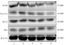
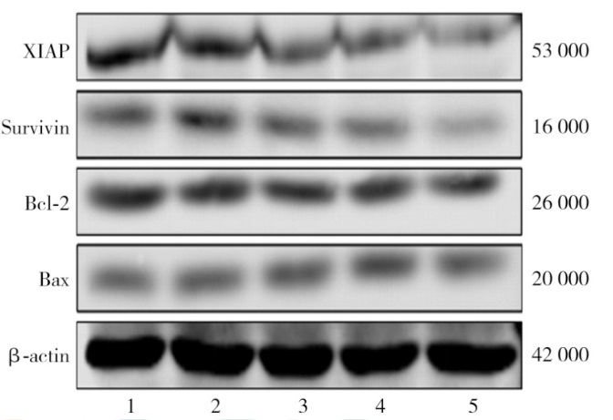
| 组别 | XIAP | Survivin | Bcl-2 | Bax |
|---|---|---|---|---|
| 阴性对照组 | 1.00±0.04 | 0.97±0.01 | 0.99±0.01 | 1.00±0.03 |
| HLY78组 | 1.01±0.08a | 1.17±0.15a | 1.23±0.10a | 0.94±0.03a |
| OA组 | 0.69±0.10ab | 0.75±0.08ab | 0.69±0.07ab | 1.32±0.09ab |
| HLY78+OA组 | 0.88±0.04abc | 0.91±0.14abc | 0.90±0.11abc | 1.11±0.02abc |
| IWR-1组 | 0.57±0.09d | 0.39±0.05d | 0.49±0.04d | 1.24±0.08d |
| F值 | 18.10 | 24.95 | 41.82 | 23.96 |
| P值 | <0.001 | <0.001 | <0.001 | <0.001 |
"
| [1] |
Zheng YY, Li LF, Shen ZB, et al. Mechanisms of neural infiltration-mediated tumor metabolic reprogramming impacting immunotherapy efficacy in non-small cell lung cancer[J]. J Exp Clin Cancer Res, 2024, 43: 284. DOI: 10.1186/s13046-024-03202-9.
pmid: 39385213 |
| [2] | 张旭旭, 李家贺, 张继朋, 等. 2025年美国临床肿瘤学会(ASCO)会议EGFR突变型非小细胞肺癌研究进展解读[J]. 中国胸心血管外科临床杂志, 2026, 33(1): 19-29. DOI: 10.7507/1007-4848.202508039. |
| [3] | Garg P, Singhal S, Kulkarni P, et al. Advances in non-small cell lung cancer: current insights and future directions[J]. J Clin Med, 2024, 13(14): 4189. DOI: 10.3390/jcm13144189. |
| [4] |
Kahn M. Can we safely target the WNT pathway?[J]. Nat Rev Drug Discov, 2014, 13(7): 513-532. DOI: 10.1038/nrd4233.
pmid: 24981364 |
| [5] | Tammela T, Sanchez-Rivera FJ, Cetinbas NM, et al. A Wnt-producing niche drives proliferative potential and progression in lung adenocarcinoma[J]. Nature, 2017, 545(7654): 355-359. DOI: 10.1038/nature22334. |
| [6] | 谷彤彤, 陈果, 刘黎明, 等. 齐墩果酸C-3及C-28位衍生物的设计合成及抗肿瘤活性研究[J]. 化学试剂, 2024, 46(10): 91-98. DOI: 10.13822/j.cnki.hxsj.2024.0050. |
| [7] | Jiang XY, Xu ZY, Wang H, et al. Metabolomics and integrated network pharmacology analysis revealed multi-targeted anti-cancer effect of FuZhengXiaoJi decoction against non-small cell lung cancer[J]. Phytomedicine, 2025, 143: 156855. DOI: 10.1016/j.phymed.2025.156855. |
| [8] |
Chu P, Li H, Luo R, et al. Oleanolic acid derivative SZC014 inhibit cell proliferation and induce apoptosis of human breast cancer cells in a ROS-dependent way[J]. Neoplasma, 2017, 64(5): 681-692. DOI: 10.4149/neo_2017_505.
pmid: 28592114 |
| [9] | Zhou WP, Zeng XJ, Wu XP. Effect of oleanolic acid on apoptosis and autophagy of SMMC-7721 hepatoma cells[J]. Med Sci Monit, 2020, 26: e921606. DOI: 10.12659/MSM.921606. |
| [10] | Edathara PM, Chintalapally S, Makani VKK, et al. Inhibitory role of oleanolic acid and esculetin in HeLa cells involve multiple signaling pathways[J]. Gene, 2021, 771: 145370. DOI: 10.1016/j.gene.2020.145370. |
| [11] | 李开瑞, 刘洁, 李小娜, 等. 齐墩果酸对肺腺癌A549细胞增殖及凋亡的影响及机制[J]. 中国医药导报, 2019, 16(31): 4-9, 181. |
| [12] | Han SH, Han JH, Chun WJ, et al. Nobiletin inhibits non-small-cell lung cancer by inactivating WNT/β-catenin signaling through downregulating miR-15-5p[J]. Evid Based Complement Alternat Med, 2021, 2021: 7782963. DOI: 10.1155/2021/7782963. |
| [13] |
Zhao X, Liu M, Li D. Oleanolic acid suppresses the proliferation of lung carcinoma cells by miR-122/cyclin G1/MEF2D axis[J]. Mol Cell Biochem, 2015, 400(1-2): 1-7. DOI: 10.1007/s11010-014-2228-7.
pmid: 25472877 |
| [14] | Lúcio KA, da Graça Rocha G, Monção-Ribeiro LC, et al. Oleanolic acid initiates apoptosis in non-small cell lung cancer cell lines and reduces metastasis of a B16F10 melanoma model in vivo[J]. PLoS One, 2011, 6(12): e28596. DOI: 10.1371/journal.pone.0028596. |
| Viewed | ||||||
|
Full text |
|
|||||
|
Abstract |
|
|||||