国际肿瘤学杂志 ›› 2026, Vol. 53 ›› Issue (6): 321-330.doi: 10.3760/cma.j.cn371439-20251128-00053

• 论著 • 上一篇    下一篇

Wnt/β-catenin信号通路在齐墩果酸调控NSCLC A549细胞增殖、凋亡中的作用与分子机制

王俊淇1,2, 李开瑞1, 袁茸1,2, 路颖1,2, 徐朝军2,3, 宋岚1,2()   

  1. 1 湖南中医药大学医学院长沙 410208
    2 湖南中医药大学湖南省血管生物学与转化医学重点实验室长沙 410208
    3 湖南中医药大学第一附属医院胸心血管外科长沙 410007
  • 收稿日期:2025-11-28 出版日期:2026-06-08 发布日期:2026-06-05
  • 通讯作者: 宋岚,Email:songlan311492@hnucm.edu.cn
  • 基金资助:
    湖南省自然科学基金(2025JJ80944);湖南省教育厅科学研究项目(22A0243);湖南省研究生创新项目(LXBZZ2024168)

Role and molecular mechanism of the Wnt/β-catenin signaling pathway in the regulation of NSCLC A549 cell proliferation and apoptosis by oleanolic acid

Wang Junqi1,2, Li Kairui1, Yuan Rong1,2, Lu Ying1,2, Xu Zhaojun2,3, Song Lan1,2()   

  1. 1 Medicine SchoolHunan University of Chinese MedicineChangsha 410208, China
    2 Hunan Provincial Key Laboratory of Vascular Biology and Translational MedicineHunan University of Chinese MedicineChangsha 410208, China
    3 Department of Cardiothoracic SurgeryFirst Hospital of Hunan University of Chinese MedicineChangsha 410007, China
  • Received:2025-11-28 Online:2026-06-08 Published:2026-06-05
  • Contact: Song Lan,Email:songlan311492@hnucm.edu.cn
  • Supported by:
    Natural Science Foundation of Hunan Province of China(2025JJ80944);Science Research Project of the Education Department of Hunan Province of China(22A0243);Innovation Project for Postgraduates of Hunan Province of China(LXBZZ2024168)

摘要:

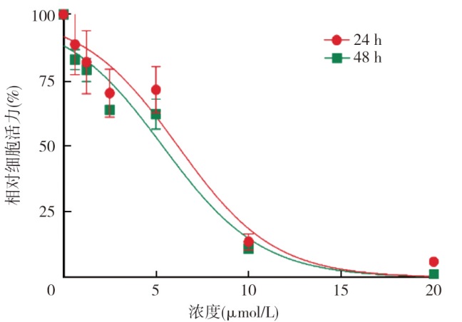
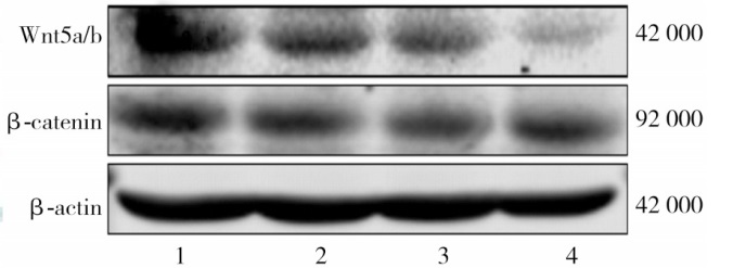
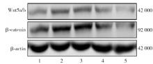
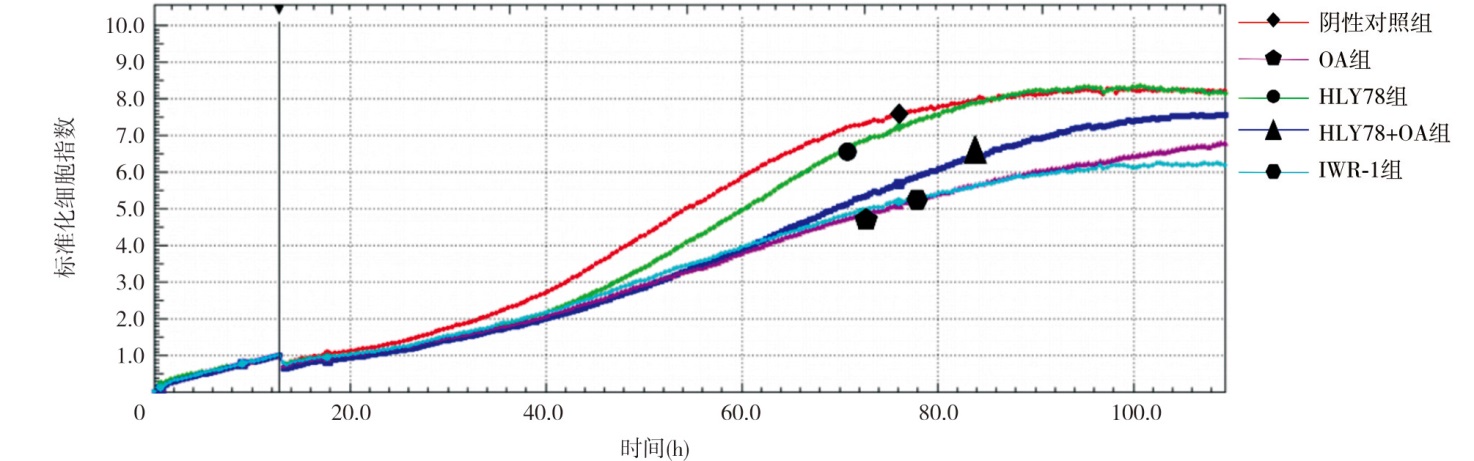
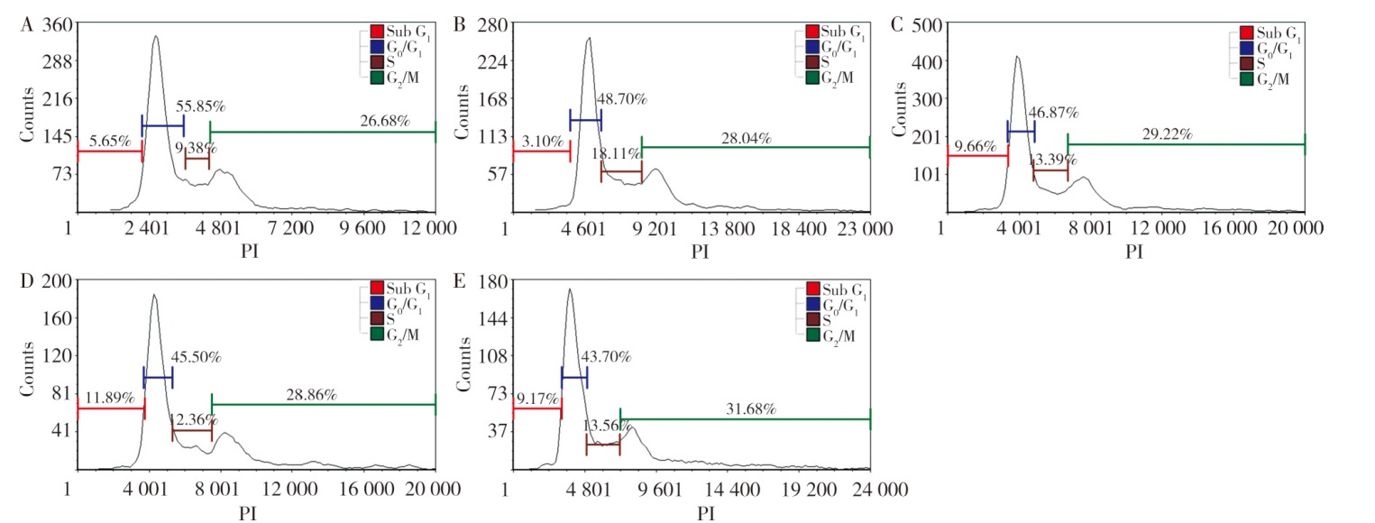
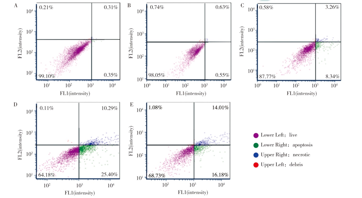
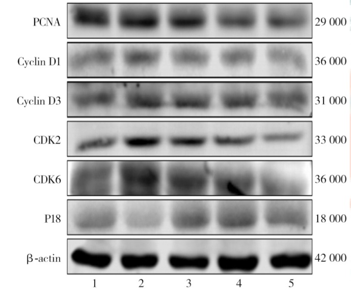
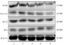
目的 探讨Wnt/β-catenin信号通路在齐墩果酸(OA)调控非小细胞肺癌(NSCLC)A549细胞增殖、凋亡中的作用及潜在的分子机制。方法 体外培养A549细胞, 采用CCK-8法检测不同浓度(0、0.625、1.25、2.5、5、10、20 μmol/L)OA对细胞活力的影响。将细胞分为阴性对照组、HLY78组(10 μmol/L HLY78)、OA组(5 μmol/L OA)、HLY78+OA组(10 μmol/L HLY78+5 μmol/L OA)、IWR-1组(8 μmol/L IWR-1)。采用蛋白质印迹法检测Wnt/β-catenin信号通路、细胞周期与凋亡相关蛋白表达水平;采用实时细胞分析技术检测细胞增殖动态;采用流式细胞术检测细胞周期和凋亡情况。结果 OA对NSCLC A549细胞活力的抑制大体呈显著的浓度和时间依赖性。阴性对照组、HLY78组、OA组、HLY78+OA组、IWR-1组NSCLC A549细胞的Wnt5a/b蛋白表达水平分别为1.03±0.06、1.42±0.02、0.98±0.07、1.17±0.05、0.34±0.10, β-catenin蛋白表达水平分别为0.99±0.02、1.22±0.08、0.69±0.16、0.85±0.12、0.39±0.19, 差异均有统计学意义(F=116.70, P<0.001;F=19.20, P<0.001);与阴性对照组相比, OA组中Wnt5a/b、β-catenin蛋白表达均显著降低(均P<0.05), 而HLY78组中Wnt5a/b、β-catenin蛋白表达均显著升高(均P<0.05);与OA组相比, HLY78+OA组中Wnt5a/b和β-catenin蛋白表达水平均显著升高(均P<0.05)。5组细胞的72 h标准化细胞指数分别为7.20±0.04、6.75±0.05、4.74±0.03、5.22±0.04、4.87±0.03, 差异有统计学意义(F=8 745.26, P<0.001);与阴性对照组相比, OA组和IWR-1组的标准化细胞指数均显著降低(均P<0.05);与OA组相比, HLY78+OA组的标准化细胞指数显著升高(P<0.05)。5组细胞周期Sub G1期占比分别为(5.45±0.21)%、(3.30±0.20)%、(9.69±1.91)%、(8.29±1.16)%、(8.21±1.26)%, G0/G1期占比分别为(55.46±0.37)%、(45.29±3.34)%、(45.07±0.42)%、(46.15±2.61)%、(52.15±4.93)%, S期占比分别为(10.81±1.24)%、(18.53±0.92)%、(13.80±2.09)%、(15.17±1.53)%、(11.73±2.76)%, 差异均有统计学意义(F=15.16, P<0.001;F=7.86, P=0.004;F=8.36, P=0.003);OA组与阴性对照组相比, HLY78+OA组与OA组及阴性对照组相比, 差异均有统计学意义(均P<0.05)。5组细胞凋亡率分别为(0.61±0.05)%、(0.90±0.24)%、(32.96±2.39)%、(13.02±3.91)%、(30.65±0.94)%, 差异有统计学意义(F=166.60, P<0.001);OA组A549凋亡率较阴性对照组升高(P<0.05);HLY78+OA组凋亡率较OA组降低(P<0.05)。5组细胞周期相关蛋白PCNA、Cyclin D1、Cyclin D3、CDK2、CDK6、P18, 凋亡相关蛋白XIAP、Survivin、Bcl-2、Bax表达差异均有统计学意义(F=20.23, P<0.001;F=17.19, P<0.001;F=63.77, P<0.001;F=24.62, P<0.001;F=127.25, P<0.001;F=11.89, P<0.001;F=18.10, P<0.001;F=24.95, P<0.001;F=41.82, P<0.001;F=23.96, P<0.001)。与阴性对照组相比, OA组中PCNA、Cyclin D1、Cyclin D3、CDK2、CDK6、XIAP、Survivin、Bcl-2 蛋白表达水平均较低, P18、Bax蛋白表达水平均较高(均P<0.05);与OA组相比, HLY78+OA组中PCNA、Cyclin D1、Cyclin D3、CDK2、CDK6、XIAP、Survivin、Bcl-2蛋白表达水平均较高, P18、Bax表达水平均较低(均P<0.05)。结论 Wnt/β-catenin信号通路在OA抑制NSCLC A549细胞增殖及诱导凋亡中发挥关键介导作用, OA主要通过靶向抑制Wnt/β-catenin信号通路, 下调细胞周期驱动蛋白及抗凋亡蛋白表达, 同时上调促凋亡蛋白表达, 从而实现抑制细胞增殖、阻滞细胞周期并促进凋亡的抗肿瘤效应。

关键词: 癌, 非小细胞肺, 齐墩果酸, 细胞增殖, 细胞凋亡

Abstract:

Objective To investigate the role of the Wnt/β-catenin signaling pathway in regulating the proliferation and apoptosis of non-small cell lung cancer(NSCLC)A549 cells by oleanolic acid(OA)and its potential molecular mechanism. Methods A549 cells were cultured in vitro, and the effect of different concentrations(0, 0.625, 1.25, 2.5, 5, 10, 20 μmol/L)of OA on cell viability was detected by CCK-8 assay. The cells were divided into the negative control group, the HLY78 group(10 μmol/L HLY78), the OA group(5 μmol/L OA), the HLY78+OA group(10 μmol/L HLY78+5 μmol/L OA), and the IWR-1 group(8 μmol/L IWR-1). Western blotting was used to detect the expression levels of proteins related to the Wnt/β-catenin signaling pathway, cell cycle, and apoptosis. Real-time cell analysis was employed to detect cell proliferation dynamics. Flow cytometry was performed to analyze the cell cycle and apoptosis. Results OA inhibited the viability of NSCLC A549 cells in a generally significant dose- and time-dependent manner. The expression levels of Wnt5a/b protein in NSCLC A549 cells of the negative control group, the HLY78 group, the OA group, the HLY78+OA group, and the IWR-1 group were 1.03±0.06, 1.42±0.02, 0.98±0.07, 1.17±0.05, and 0.34±0.10, respectively, and the expression levels of β-catenin protein were 0.99±0.02, 1.22±0.08, 0.69±0.16, 0.85±0.12, and 0.39±0.19, respectively, with statistically significant differences(F=116.70, P<0.001; F=19.20, P<0.001). Compared with the negative control group, the protein expressions of Wnt5a/b and β-catenin in the OA group were both significantly decreased(both P<0.05), while the protein expressions of Wnt5a/b and β-catenin in the HLY78 group were significantly increased(both P<0.05). Compared with the OA group, the protein expressions of Wnt5a/b and β-catenin in the HLY78+OA group were both significantly increased(both P<0.05). The 72 h normalized cell indexes of the 5 groups were 7.20±0.04, 6.75±0.05, 4.74±0.03, 5.22±0.04, and 4.87±0.03, respectively, with a statistically significant difference(F=8 745.26, P<0.001). Compared with the negative control group, the normalized cell indexes of the OA group and the IWR-1 group were both significantly decreased(both P<0.05); compared with the OA group, the normalized cell index of the HLY78+OA group was significantly increased(P<0.05). The proportions of Sub G1 phase in the 5 groups were(5.45±0.21)%, (3.30±0.20)%, (9.69±1.91)%, (8.29±1.16)%, and(8.21±1.26)%, respectively; the proportions of G0/G1 phase were(55.46±0.37)%, (45.29±3.34)%, (45.07±0.42)%, (46.15±2.61)%, and(52.15±4.93)%, respectively; the proportions of S phase were(10.81±1.24)%, (18.53±0.92)%, (13.80±2.09)%, (15.17±1.53)%, and(11.73±2.76)%, respectively, and with statistically significant differences(F=15.16, P<0.001; F=7.86, P=0.004; F=8.36, P=0.003). Compared with the negative control group, the OA group showed statistically significant differences. Additionally, compared with both the OA group and the negative control group, the HLY78+OA group also demonstrated statistically significant differences(all P<0.05). The apoptosis rates of the 5 groups were(0.61±0.05)%, (0.90±0.24)%, (32.96±2.39)%, (13.02±3.91)%, and(30.65±0.94)%, respectively, with a statistically significant difference(F=166.60, P<0.001). The apoptosis rate of A549 cells in the OA group was significantly increased compared with the negative control group(P<0.05); the apoptosis rate in the HLY78+OA group was significantly decreased compared with the OA group(P<0.05). There were statistically significant differences in the expression levels of cell cycle-related proteins(PCNA, Cyclin D1, Cyclin D3, CDK2, CDK6, P18)and apoptosis-related proteins(XIAP, Survivin, Bcl-2, Bax)among the 5 groups(F=20.23, P<0.001; F=17.19, P<0.001; F=63.77, P<0.001; F=24.62, P<0.001; F=127.25, P<0.001; F=11.89, P<0.001; F=18.10, P<0.001; F=24.95, P<0.001; F=41.82, P<0.001; F=23.96, P<0.001). Compared with the negative control group, the protein expressions of PCNA, Cyclin D1, Cyclin D3, CDK2, CDK6, XIAP, Survivin, Bcl-2 in the OA group were all significantly lower, while the protein expressions of P18 and Bax were higher(all P<0.05). Compared with the OA group, the protein expressions of PCNA, Cyclin D1, Cyclin D3, CDK2, CDK6, XIAP, Survivin, Bcl-2 in the HLY78+OA group were all higher, while the expressions of P18 and Bax were lower(all P<0.05). Conclusions The Wnt/β-catenin signaling pathway critically mediates the anti-tumor effects of OA in NSCLC A549 cells. By targeting and inhibiting this pathway, OA downregulates cell cycle drivers and anti-apoptotic proteins while upregulating pro-apoptotic proteins, thereby effectively inhibiting proliferation, arresting the cell cycle, and promoting apoptosis.

Key words: Carcinoma, non-small-cell lung, Oleanolic acid, Cell proliferation, Apoptosis