国际肿瘤学杂志 ›› 2026, Vol. 53 ›› Issue (6): 346-354.doi: 10.3760/cma.j.cn371439-20251123-00056

• 论著 • 上一篇    下一篇

NSCLC影像组学特征与临床特征构建的联合模型对其骨转移的预测效能研究

李佳凝1, 姚学敏1, 王金云2, 贾敬好1(), 孙国贵1()   

  1. 1 华北理工大学附属医院肿瘤放化疗科唐山 063000
    2 解放军总医院第四医学中心胸外科北京 100036
  • 收稿日期:2025-11-23 出版日期:2026-06-08 发布日期:2026-06-05
  • 通讯作者: 贾敬好,Email:jjh0322@163.com
    孙国贵,Email:guogui_sun2021@sina.com
  • 基金资助:
    国家自然科学基金(82172658);河北省医学科学研究课题(20191595)

Predictive efficacy of a combined model of radiomics features and clinical signatures for bone metastasis in NSCLC

Li Jianing1, Yao Xuemin1, Wang Jinyun2, Jia Jinghao1(), Sun Guogui1()   

  1. 1 Department of Oncology Radiotherapy and ChemotherapyNorth China University of Science and Technology Affiliated HospitalTangshan 063000, China
    2 Department of Thoracic SurgeryFourth Medical Center of Chinese PLA General HospitalBeijing 100036, China
  • Received:2025-11-23 Online:2026-06-08 Published:2026-06-05
  • Contact: Jia Jinghao,Email:jjh0322@163.com
    Sun Guogui,Email:guogui_sun2021@sina.com
  • Supported by:
    National Natural Science Foundation of China(82172658);Medical Science Research Project of Hebei Province of China(20191595)

摘要:

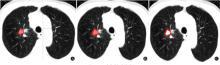
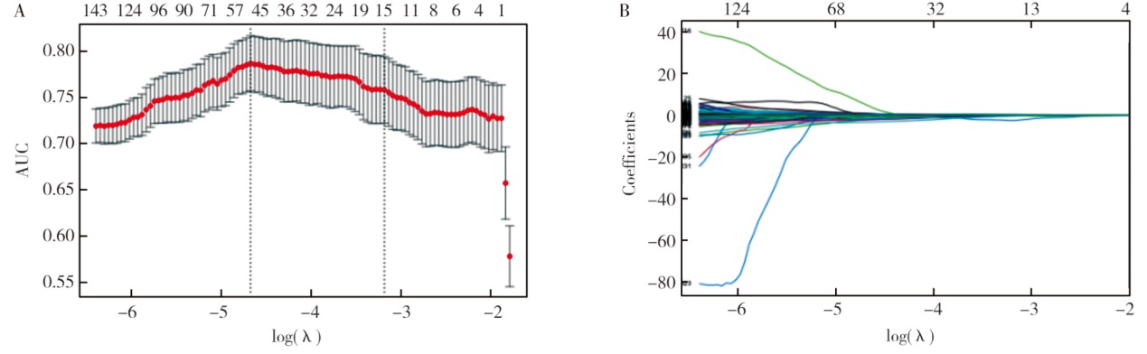
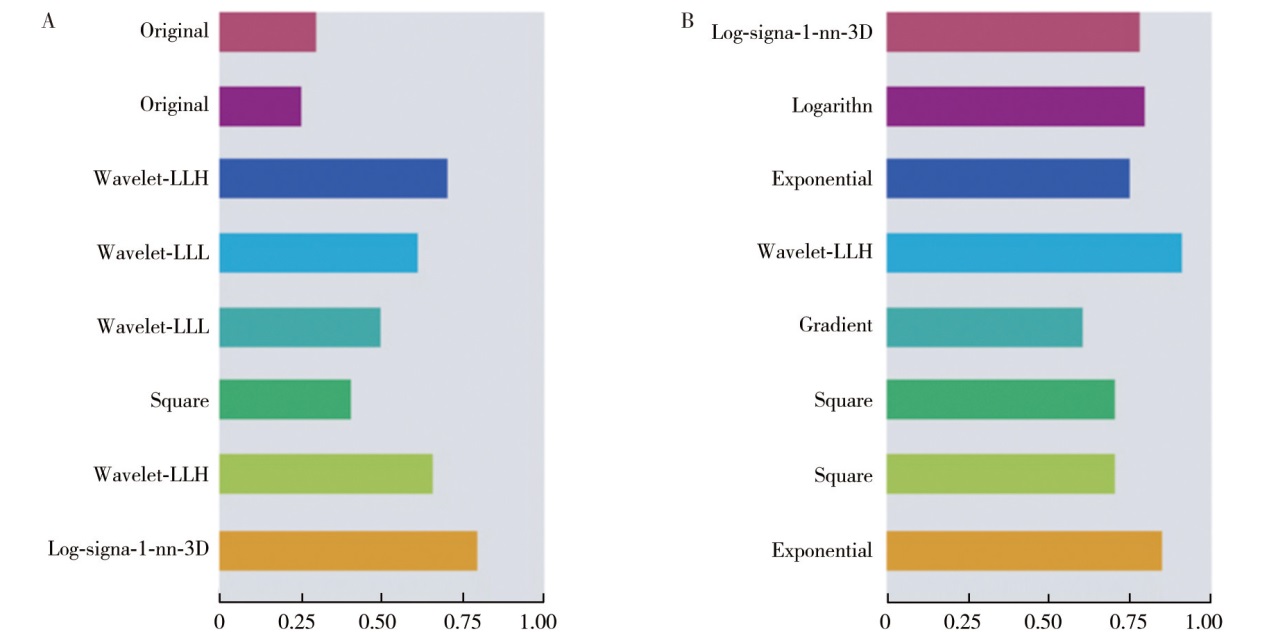
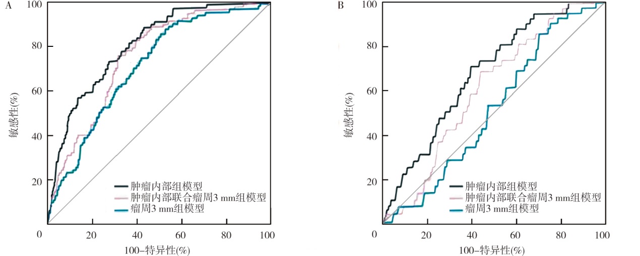
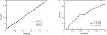
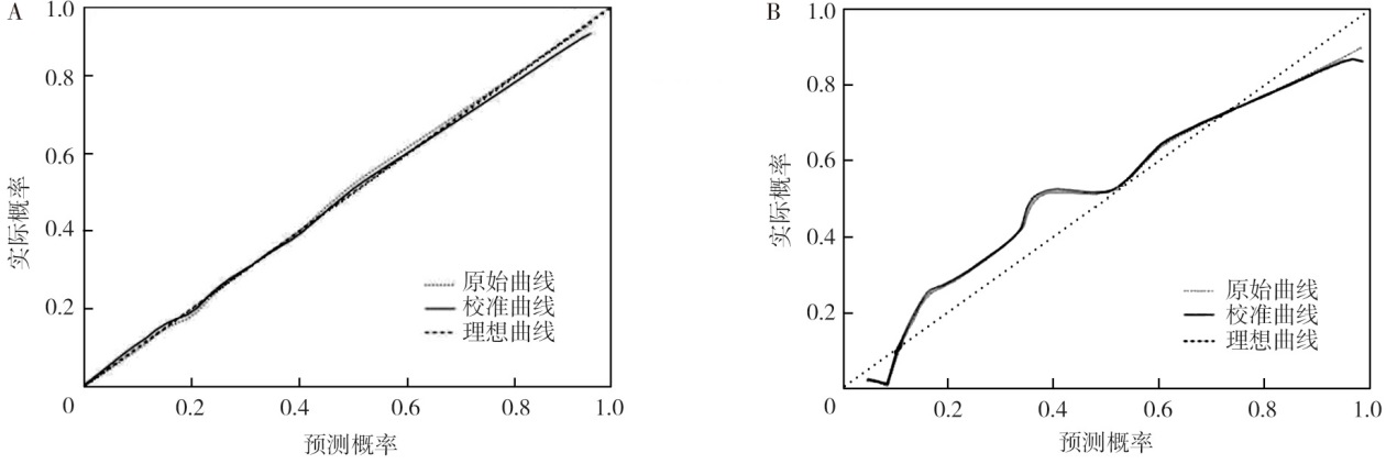
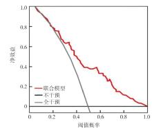
目的 探讨瘤内联合瘤周组织的CT影像组学特征与临床特征构建的联合模型对非小细胞肺癌(NSCLC)骨转移的预测效能。方法 回顾性分析唐山市人民医院2017年1月至2019年12月经病理证实的537例NSCLC患者的胸部动脉期CT图像及临床资料, 按照7∶3比例将患者分为训练集(n=376)和验证集(n=161)。在训练集中构建预测模型, 在训练集和验证集中分别对模型进行预测效能评估和临床应用价值验证。采用单因素及多因素logistic回归分析NSCLC患者发生骨转移的影响因素。构建肿瘤内部组、肿瘤内部联合瘤周3 mm组、单纯瘤周3 mm组的影像组学模型, 并选出最优模型联合临床特征构建联合模型。采用受试者操作特征(ROC)曲线、校准曲线及临床决策曲线分析(DCA)评估模型的诊断效能及临床应用价值。结果 537例NSCLC患者中发生骨转移414例, 其中训练集290例、验证集124例。单因素分析显示, 吸烟史、肿瘤位置、T分期、N分期、病理类型、D-二聚体、癌胚抗原(CEA)、细胞角蛋白19片段抗原21-1、鳞状细胞癌相关抗原、毛刺征、分叶征、胸膜凹陷征、血管集束征均是预测NSCLC患者发生骨转移的影响因素(均P<0.05)。多因素分析显示, T分期(OR=0.69, 95%CI为0.52~0.87, P<0.001)、N分期(OR=0.24, 95%CI为0.13~0.43, P<0.001)、病理类型(OR=6.01, 95%CI为2.83~12.77, P<0.001)、D-二聚体(OR=0.32, 95%CI为0.17~0.59, P<0.001)、CEA(OR=0.25, 95%CI为0.14~0.44, P<0.001)、毛刺征(OR=0.21, 95%CI为0.07~0.65, P=0.007)、胸膜凹陷征(OR=0.32, 95%CI为0.18~0.56, P<0.001)均是预测NSCLC患者发生骨转移的独立影响因素。ROC曲线分析显示, 肿瘤内部组模型、肿瘤内部联合瘤周3 mm组模型、瘤周3 mm组模型预测NSCLC患者发生骨转移的曲线下面积(AUC)在训练集中分别为0.81、0.79、0.74, 肿瘤内部组模型的预测价值高于肿瘤内部联合瘤周3 mm组模型、瘤周3 mm组模型(Z=1.46, P=0.032;Z=3.01, P=0.024);验证集中的AUC分别为0.66、0.63、0.53, 肿瘤内部组模型的预测价值高于肿瘤内部联合瘤周3 mm组模型、瘤周3 mm组模型(Z=2.37, P=0.025;Z=4.12, P=0.012)。选择肿瘤内部组影像组学和多因素分析中有统计学意义的影响因素构建联合模型, 训练集中联合模型的AUC为0.94, 预测价值高于肿瘤内部组模型(Z=2.43, P=0.023);验证集中联合模型的AUC为0.92, 预测价值高于肿瘤内部组模型(Z=3.76, P=0.007)。校准曲线显示, 训练集和验证集实际发生概率均与预测概率较为一致。DCA显示, 联合模型的辨别能力较好。结论 肿瘤T分期、N分期、病理类型、D-二聚体、CEA、毛刺征、胸膜凹陷征均是预测NSCLC患者发生骨转移的独立影响因素;在影像组学模型中, 肿瘤内部组的影像组学模型具有较高的预测NSCLC骨转移效能;基于上述因素构建的联合模型可进一步提高NSCLC骨转移的预测效能, 具有潜在的临床应用价值。

关键词: 癌, 非小细胞肺, 肿瘤转移, 影像组学, 胸部增强CT, 预测模型

Abstract:

Objective To explore the predictive efficacy of a combined model integrating intratumoral and peritumoral CT radiomic features and clinical signatures for bone metastasis in non-small cell lung cancer(NSCLC). Methods A retrospective analysis was conducted on chest arterial-phase CT images and clinical data from 537 pathologically confirmed NSCLC patients at Tangshan People's Hospital between January 2017 and December 2019. Patients were divided into a training cohort(n=376)and a validation set(n=161)in a 7∶3 ratio. Predictive models were developed within the training set, and the predictive efficacy was evaluated in both the training and validation sets respectively, and the clinical application value was verified as well. Using univariate and multivariate logistic regression analysis, the influencing factors for bone metastasis in NSCLC patients were investigated. Radiomics models were established for the tumor interior group, the tumor interior combined with a 3 mm peritumoral group, and the simple 3 mm peritumoral group. The best model was selected and combined with clinical signatures to construct a combined model. The diagnostic efficacy and clinical application value of the model were evaluated using receiver operator characteristic(ROC)curves, the calibration curve, and decision curve analysis(DCA). Results Among the 537 NSCLC patients, 414 had bone metastasis(290 in training set, 124 in validation set). Univariate analysis showed that, smoking history, tumor location, T stage, N stage, pathological type, D-dimer, carcinoembryonic antigen(CEA), cytokeratin fragment antigen 21-1, squamous cell carcinoma antigen, spiculation sign, lobulation sign, pleural indentation sign, and vascular convergence sign were all influencing factors predicting bone metastasis in NSCLC patients(all P<0.05). Multivariate analysis showed that, T stage(OR=0.69, 95%CI:0.52-0.87, P<0.001), N stage(OR=0.24, 95%CI:0.13-0.43, P<0.001), pathological type(OR=6.01, 95%CI:2.83-12.77, P<0.001), D-dimer(OR=0.32, 95%CI:0.17-0.59, P<0.001), CEA(OR=0.25, 95%CI:0.14-0.44, P<0.001), spiculation sign(OR=0.21, 95%CI:0.07-0.65, P=0.007), and pleural indentation sign(OR=0.32, 95%CI:0.18-0.56, P<0.001)were all independent influencing factors predicting bone metastasis in NSCLC patients. ROC curve analysis showed that, the area under the curve(AUC)for predicting bone metastasis in the training set were 0.81, 0.79, and 0.74 for the models in the tumor interior group, the tumor interior combined with a 3 mm peritumoral group, and the simple 3 mm peritumoral group, respectively. The predictive value of the model in the tumor interior group was higher than that of the models in the tumor interior combined with a 3 mm peritumoral group and the simple 3 mm peritumoral group(Z=1.46, P=0.032; Z=3.01, P=0.024). In the validation set, the AUC were 0.66, 0.63, and 0.53, respectively, with the predictive value of the model in the tumor interior group being higher than that of the models in the tumor interior combined with a 3 mm peritumoral group and the simple 3 mm peritumoral group(Z=2.37, P=0.025; Z=4.12, P=0.012). A combined model was established using the radiomic features in the tumor interior group and the influencing factors with statistical significance from the multivariate analysis. The AUC of the combined model was 0.94 in the training set, which was higher than that of the model in the tumor interior group alone(Z=2.43, P=0.023). In the validation set, the combined model's AUC was 0.92, which was also higher than that of the model in the tumor interior group(Z=3.76, P=0.007). The calibration curve showed that the actual probabilities of both the training set and the validation set were in relatively good agreement with the predicted probabilities. DCA showed good discrimination ability for the combined model. Conclusions T stage, N stage, pathological type, D-dimer, CEA, spiculation sign, pleural indentation sign are all independent influencing factors predicting bone metastasis in NSCLC patients. Among the radiomics models, the model in the tumor interior group demonstrates higher predictive efficacy for NSCLC bone metastasis. The combined model constructed based on the above factors can further improve the predictive efficacy of bone metastasis in NSCLC patients,showing promising potential for clinical application.

Key words: Carcinoma, non-small-cell lung, Neoplasm metastasis, Radiomics, Chest-enhanced computed tomography, Prediction model