国际肿瘤学杂志 ›› 2025, Vol. 52 ›› Issue (9): 554-559.doi: 10.3760/cma.j.cn371439-20250530-00094

• 论著 • 上一篇    下一篇

基于kV正交图像引导的头部肿瘤伽玛刀无痛面模分次治疗的摆位误差分析研究

李鹏1, 张双1, 刘华锋2, 纪娜2, 候向坤2, 席奥航2, 宗建海2()   

  1. 1西安国际医学中心医院放射治疗科,西安 710100
    2西安国际医学中心医院伽玛刀治疗中心,西安 710100
  • 收稿日期:2025-05-30 修回日期:2025-07-20 出版日期:2025-09-08 发布日期:2025-10-21
  • 通讯作者: 宗建海 E-mail:1911688816@qq.com

Research on positioning errors analysis of gamma knife pain-free face mask fractionated treatment for head tumors based on kV orthogonal image guidance

Li Peng1, Zhang Shuang1, Liu Huafeng2, Ji Na2, Hou Xiangkun2, Xi Aohang2, Zong Jianhai2()   

  1. 1Radiation Therapy Center, Xi'an International Medical Center Hospital, Xi'an 710100, China
    2Gamma Knife Treatment Center, Xi'an International Medical Center Hospital, Xi'an 710100, China
  • Received:2025-05-30 Revised:2025-07-20 Online:2025-09-08 Published:2025-10-21
  • Contact: Zong Jianhai E-mail:1911688816@qq.com

摘要:

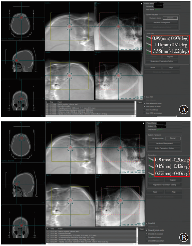
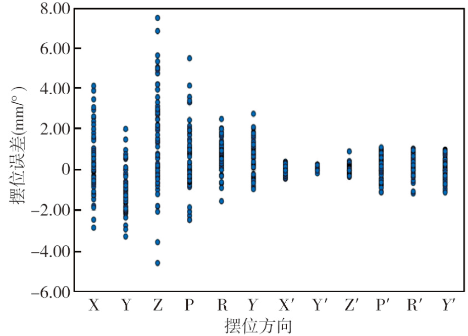
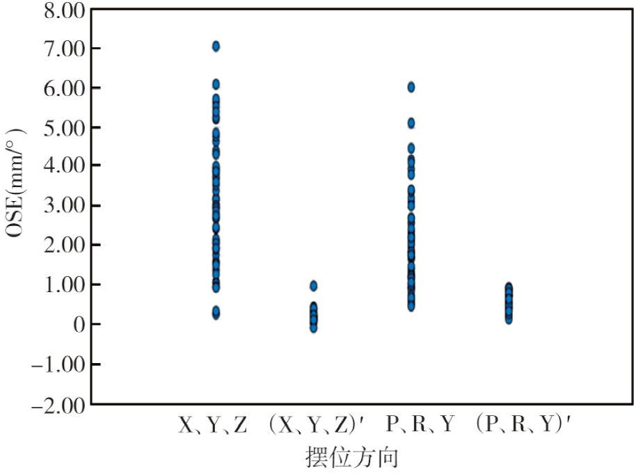
目的 分析基于kV正交图像引导的头部肿瘤伽玛刀无痛面模分次治疗患者摆位误差和综合摆位误差(OSE)。方法 选取2022年7月1日至2024年5月31日西安国际医学中心医院伽玛刀治疗中心收治的58例行头部肿瘤图像引导伽玛刀无痛面模分次治疗的患者作为研究对象。通过kV级正交X射线IGPS图像引导定位系统,采集患者校正前左右(X)、前后(Y)、头脚(Z)3个平移方向和左右(P)、前后(R)、头脚(Y)3个旋转方向摆位误差,经在线校正并结合人工摆位复核验证,再次计算获得校正后摆位误差。计算平移和旋转方向校正前、后OSE。绘制校正前、后X、Y、Z、P、R、Y 6个方向的摆位误差以及平移和旋转方向OSE散点图。比较6个方向校正前、后的摆位误差以及平移和旋转方向OSE,并比较不同年龄段、性别患者平移及旋转方向的OSE。结果 患者X、Y、Z、P、R、Y 6个方向上的校正前摆位误差分别为(0.45±1.54)mm、-0.96(-1.70,-0.28)mm、1.67(-0.15,3.07)mm、(0.70±1.60)°、0.65(0.30,1.19)°、(0.59±0.87)°,校正后分别为(-0.02±0.18)mm、0.15(0.10,0.21)mm、0.06(-0.04,0.16)mm、(0.20±0.79)°、0.42(0.19,0.78)°、(0.20±0.63)°,校正前、后差异均有统计学意义(t=2.30,P=0.025;Z=-5.43,P<0.001;Z=-4.10,P<0.001;t=2.56,P=0.013;Z=-3.21,P=0.001;t=3.21,P=0.002)。平移(X、Y、Z)和旋转(P、R、Y)方向校正前OSE分别为3.07(1.93,4.35)mm、1.90(1.28,2.66)°,校正后分别为0.27(0.21,0.33)mm、1.08(0.70,1.54)°,校正前、后差异均有统计学意义(Z=-6.60,P<0.001;Z=-5.52,P<0.001)。18~44岁年龄段患者平移(X、Y、Z)和旋转(P、R、Y)方向校正前、后OSE分别为3.65(1.62,3.95)、0.21(0.21,0.31)mm,3.25(2.24,3.96)°、0.92(0.59,1.45)°;45~59岁年龄段分别为3.57(2.17,5.22)、0.29(0.22,0.35)mm,1.89(1.30,2.30)°、1.08(0.62,1.51)°;60~74岁年龄段分别为2.92(1.74,4.06)、0.24(0.19,0.35)mm,2.16(1.09,2.95)°、0.98(0.78,1.75)°;75~89岁年龄段分别为3.24(2.12,4.37)、0.29(0.22,0.47)mm,1.73(1.01,1.83)°、0.60(0.47,1.51)°;4个年龄段间平移、旋转方向校正前、后OSE差异均无统计学意义(H=1.23,P=0.747;H=1.74,P=0.627;H=7.45,P=0.059;H=2.80,P=0.424)。男性患者平移(X、Y、Z)及旋转(P、R、Y)方向校正前、后OSE分别为(3.19±1.59)、0.27(0.27,0.33)mm,1.89(1.27,2.75)°、(0.84±0.59)°;女性患者分别为(3.22±1.99)、0.26(0.25,0.35)mm,1.90(1.34,2.41)°、(1.04±0.46)°;男、女性别间平移、旋转方向校正前、后OSE差异均无统计学意义(t=-0.07,P=0.949;Z=-0.48,P=0.632;Z=-0.02,P=0.161;t=-2.80,P=0.424)。结论 基于kV正交X射线立体成像图像引导系统,通过“自动校准+人工复核”双重验证引导,可明显减少头部肿瘤患者伽玛刀无痛面模分次治疗间的摆位误差,提高伽玛刀的摆位精度。

关键词: 头颈部肿瘤, 放射外科手术, 伽玛刀, 无痛面模, 摆位误差

Abstract:

Objective To analyze the positioning error and the overall setup errors (OSEs) of patients undergoing gamma knife pain-free face mask fractionated treatment for head tumors based on kV orthogonal image guidance. Methods A total of 58 patients who received image-guided fractionated gamma knife treatment for head tumors with a pain-free face mask at the Gamma Knife Treatment Center of Xi'an International Medical Center Hospital from July 1, 2022 to May 31, 2024 were included in the study. A kV-class orthogonal X-ray IGPS image-guided positioning system was used to collect positioning errors in three translational directions: left-right (X), anterior-posterior (Y), and head-foot (Z), as well as in three rotational directions: left-right (P), anterior-posterior (R), and head-foot (Y) before correction. After online correction and combined with manual positioning verification, the corrected positioning errors were recalculated. The OSEs in translational and rotational directions were calculated before and after correction. The positioning errors in all six directions (X, Y, Z, P, R, Y) before and after correction were plotted. And the OSE scatter plots in translational and rotational directions were created accordingly. Errors in the six directions and OSEs in translational and rotational directions were compared. The OSEs in translational and rotational directions were analyzed across different age groups and genders. Results The pre-correction positioning errors in the X, Y, Z, P, R, Y directions for patients were (0.45±1.54) mm, -0.96 (-1.70, -0.28) mm, 1.67 (-0.15, 3.07) mm, (0.70±1.60)°, 0.65 (0.30, 1.19)°, (0.59±0.87)°, and the post-correction positioning errors were (-0.02±0.18) mm, 0.15 (0.10, 0.21) mm, 0.06 (-0.04, 0.16) mm, (0.20±0.79)°, 0.42 (0.19, 0.78)°, (0.20±0.63)°. There were statistically significant differences between before and after correction (t=2.30, P=0.025; Z=-5.43, P<0.001; Z=-4.10, P<0.001; t=2.56, P=0.013; Z=-3.21, P=0.001; t=3.21, P=0.002). The OSEs in translational (X, Y, Z) and rotational (P, R, Y) directions before correction were 3.07 (1.93, 4.35) mm, 1.90 (1.28, 2.66)°, and the OSEs after correction were 0.27 (0.21, 0.33) mm, 1.08 (0.70, 1.54)°, with statistically significant differences (Z=-6.60, P<0.001; Z=-5.52, P<0.001). For patients aged 18-44 years, the OSEs in translational (X, Y, Z) and rotational (P, R, Y) directions before and after correction were 3.65 (1.62, 3.95), 0.21 (0.21, 0.31) mm, 3.25 (2.24, 3.96)°, 0.92 (0.59, 1.45)°; for patients aged 45-59 years, the OSEs were 3.57 (2.17, 5.22), 0.29 (0.22, 0.35) mm, 1.89 (1.30, 2.30)°, 1.08 (0.62, 1.51)°; for patients aged 60-74 years, the OSEs were 2.92 (1.74, 4.06), 0.24 (0.19, 0.35) mm, 2.16 (1.09, 2.95)°, 0.98 (0.78, 1.75)°; for patients aged 75-89 years, the OSEs were 3.24 (2.12, 4.37), 0.29 (0.22, 0.47) mm, 1.73 (1.01, 1.83)°, 0.60 (0.47, 1.51)°. There were no statistically significant differences in OSEs of translational and rotational directions before and after correction among the four age groups (H=1.23, P=0.747; H=1.74, P=0.627; H=7.45, P=0.059; H=2.80, P=0.424). For male patients, the OSEs before and after correction in translational (X, Y, Z) and rotational (P, R, Y) directions were (3.19±1.59), 0.27 (0.27, 0.33) mm, 1.89 (1.27, 2.75)°, (0.84±0.59)°; for female patients, the OSEs were (3.22±1.99), 0.26 (0.25, 0.35) mm, 1.90 (1.34, 2.41)°, (1.04±0.46)°. There were no statistically significant differences in OSEs of translational and rotational directions before and after correction between genders (t=-0.07, P=0.949; Z=-0.48, P=0.632; Z=-0.02, P=0.161; t=-2.80, P=0.424). Conclusions The image-guided system, which is based on the kV orthogonal X-ray stereoscopic imaging, can significantly reduce the positioning errors between fractions of pain-free face mask gamma knife treatment for head tumor patients and improve the positioning accuracy of the gamma knife through the dual verification process of "automatic correction and manual review".

Key words: Head and neck neoplasms, Radiosurgery, Gamma knife, Pain-free face mask, Setup error