国际肿瘤学杂志 ›› 2025, Vol. 52 ›› Issue (9): 545-553.doi: 10.3760/cma.j.cn371439-20250704-00093

• 论著 • 上一篇    下一篇

白术内酯Ⅱ对结肠癌小鼠的抗肿瘤作用及免疫调节机制

王梦菊, 王霞()   

  1. 南京中医药大学第二附属医院 江苏省第二中医院肿瘤科,南京 210017
  • 收稿日期:2025-07-04 修回日期:2025-07-19 出版日期:2025-09-08 发布日期:2025-10-21
  • 通讯作者: 王霞 E-mail:wangxia7904@163.com
  • 基金资助:
    国家自然科学基金(82474375);南京中医药大学第二附属医院(江苏省第二中医院)院级课题(SEZJY2023015)

Anti-tumor effect and immunomodulatory mechanism of atractylenolide Ⅱ on colon cancer mice

Wang Mengju, Wang Xia()   

  1. Department of Oncology, Jiangsu Second Chinese Medicine Hospital, Second Affiliated Hospital of Nanjing University of Chinese Medicine, Nanjing 210017, China
  • Received:2025-07-04 Revised:2025-07-19 Online:2025-09-08 Published:2025-10-21
  • Contact: Wang Xia E-mail:wangxia7904@163.com
  • Supported by:
    National Natural Science Foundation of China(82474375);Hospital-Level Project of Jiangsu Second Chinese Medicine Hospital, Second Affiliated Hospital of Nanjing University of Chinese Medicine(SEZJY2023015)

摘要:

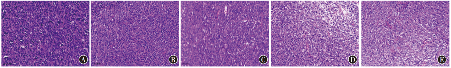
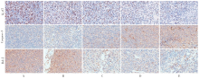
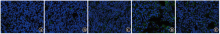
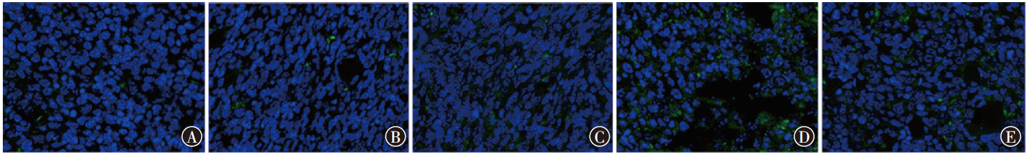
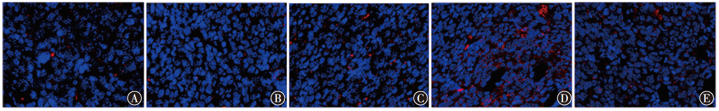
目的 探讨白术内酯Ⅱ(ATL-Ⅱ)对结肠癌的抗肿瘤作用及其免疫调节机制。方法 建立C57BL/6小鼠结肠癌皮下移植瘤模型,采用随机数字表法将小鼠随机分为模型组(PBS腹腔注射),ATL-Ⅱ低(20 mg/kg)、中(40 mg/kg)、高(60 mg/kg)剂量组及5-氟尿嘧啶(5-FU 30 mg/kg)组,每组各5只,连续给药21 d。采用HE染色检测肿瘤组织病理变化,免疫组织化学法检测肿瘤组织Ki-67、Caspase-3、Bcl-2表达水平,免疫荧光法检测肿瘤组织CD8+ T、NK1.1细胞阳性细胞率,ELISA法检测血清颗粒酶B(GzmB)、λ干扰素(IFN-λ)水平,蛋白质印迹法检测PD-L1、ERK/MAPK信号通路蛋白表达水平。结果 模型组,ATL-Ⅱ低、中、高剂量组,5-FU组结肠癌移植瘤小鼠肿瘤体积分别为(1 845.17±65.72)、(1 637.20±122.65)、(1 232.86±209.16)、(1 002.29±41.84)、(911.59±294.71)mm3,差异有统计学意义(F=125.61,P<0.001);ATL-Ⅱ低、中、高剂量组,5-FU组与模型组相比,差异均有统计学意义(均P<0.05);ATL-Ⅱ中、高剂量组,5-FU组与低剂量组相比,差异均有统计学意义(均P<0.05),且中、高剂量组间差异有统计学意义(P<0.05);5-FU组与ATL-Ⅱ中、高剂量组相比,差异均有统计学意义(均P<0.05)。5组小鼠肿瘤质量分别为(1.34±0.11)、(1.26±0.09)、(0.93±0.07)、(0.94±0.10)、(0.59±0.08)g,差异有统计学意义(F=88.88,P<0.001);ATL-Ⅱ中、高剂量组,5-FU组与模型组相比,差异均有统计学意义(均P<0.05);ATL-Ⅱ中、高剂量组,5-FU组与低剂量组相比,差异均有统计学意义(均P<0.05);5-FU组与ATL-Ⅱ中、高剂量组相比,差异均有统计学意义(均P<0.05)。5组小鼠脾脏指数分别为7.42±0.88、7.38±1.32、8.42±0.78、9.72±1.18、6.16±1.05,差异有统计学意义(F=33.20,P<0.001);ATL-Ⅱ中、高剂量组,5-FU组与模型组相比,差异均有统计学意义(均P<0.05);ATL-Ⅱ中、高剂量组,5-FU组与低剂量组相比,差异均有统计学意义(均P<0.05),且高剂量组高于中剂量组(P<0.05);5-FU组与ATL-Ⅱ中、高剂量组相比,差异均有统计学意义(均P<0.05)。HE染色结果表明,模型组小鼠肿瘤组织呈细胞密集、核大深染、异型性、细胞间质减少等典型的恶性肿瘤特征,经ATL-Ⅱ和5-FU处理的小鼠肿瘤组织中,细胞增殖显著减少,细胞排列较为疏松,核分裂现象减少,且坏死区域明显缩小,ATL-Ⅱ中、高剂量组及5-FU组能观察到较小的圆形、椭圆形细胞,核大且染色质深。5组小鼠的Ki-67、Caspase-3、Bcl-2阳性区域占比差异均有统计学意义(F=13.86,P=0.043;F=477.63,P<0.001;F=40.48,P<0.001);ATL-Ⅱ中、高剂量组,5-FU组与模型组相比,差异均有统计学意义(均P<0.05);ATL-Ⅱ高剂量组、5-FU组与低剂量组相比,差异均有统计学意义(均P<0.05)。5-FU组与ATL-Ⅱ中、高剂量组相比,Caspase-3阳性区域占比差异均有统计学意义(均P<0.05);ATL-Ⅱ中、高剂量组间差异有统计学意义(P<0.05)。5组小鼠肿瘤组织CD8+ T细胞阳性细胞率分别为(10.33±3.53)%、(15.00±5.65)%、(30.33±10.51)%、(59.33±9.04)%、(33.62±9.11)%,差异有统计学意义(F=96.33,P<0.001);ATL-Ⅱ低、中、高剂量组,5-FU组与模型组相比,差异均有统计学意义(均P<0.05);ATL-Ⅱ中、高剂量组,5-FU组与低剂量组相比,差异均有统计学意义(均P<0.05),且中、高剂量组间差异有统计学意义(P<0.05);5-FU组与ATL-Ⅱ高剂量组相比,差异有统计学意义(P<0.05)。5组小鼠肿瘤组织中NK1.1细胞阳性细胞率分别为(12.33±6.52)%、(13.00±7.00)%、(35.33±9.51)%、(43.67±12.21)%、(14.50±7.05)%,差异有统计学意义(F=283.17,P<0.001);ATL-Ⅱ中、高剂量组,5-FU组与模型组相比,差异均有统计学意义(均P<0.05);ATL-Ⅱ高剂量组与低剂量组相比,差异有统计学意义(P<0.05);5-FU组与ATL-Ⅱ中、高剂量组相比,差异均有统计学意义(均P<0.05)。5组小鼠血清GzmB水平分别为(5.00±1.00)、(5.27±0.76)、(8.27±0.61)、(10.00±1.21)、(6.15±0.69)ng/L,差异有统计学意义(F=21.45,P<0.001);ATL-Ⅱ中、高剂量组,5-FU组与模型组相比,差异均有统计学意义(均P<0.05);ATL-Ⅱ中、高剂量组与低剂量组相比,差异均有统计学意义(均P<0.05),ATL-Ⅱ中、高剂量组间差异有统计学意义(P<0.05);5-FU组与ATL-Ⅱ中、高剂量组相比,差异均有统计学意义(均P<0.05)。5组小鼠血清IFN-λ水平分别为(617.33±65.06)、(743.33±40.41)、(910.00±36.06)、(1 009.00±35.54)、(703.62±56.00)ng/L,差异有统计学意义(F=43.08,P<0.001); ATL-Ⅱ低、中、高剂量组与模型组相比,差异均有统计学意义(均P<0.05);ATL-Ⅱ中、高剂量组与低剂量组相比,差异均有统计学意义(均P<0.05),且中、高剂量组间差异有统计学意义(P<0.05);5-FU组与ATL-Ⅱ中、高剂量组相比,差异均有统计学意义(均P<0.05)。5组小鼠肿瘤组织中PD-L1、p-ERK/ERK及p-MEK/MEK蛋白表达水平差异均有统计学意义(F=125.34,P<0.001;F=89.63,P<0.001;F=35.33,P=0.002)。ATL-Ⅱ低、中、高剂量组,5-FU组与模型组相比,PD-L1表达水平差异均有统计学意义(均P<0.05); ATL-Ⅱ高剂量组、5-FU组与低剂量组相比,差异均有统计学意义(均P<0.05);ATL-Ⅱ中、高剂量组差异有统计学意义(P<0.05);ATL-Ⅱ中剂量组与5-FU组差异有统计学意义(P<0.05)。ATL-Ⅱ低、中、高剂量组,5-FU组与模型组相比,肿瘤组织中p-ERK/ERK表达水平差异均有统计学意义(均P<0.05);ATL-Ⅱ中、高剂量组,5-FU组与低剂量组相比,差异均有统计学意义(均P<0.05);5-FU组与ATL-Ⅱ中、高剂量组相比,差异均有统计学意义(均P<0.05)。ATL-Ⅱ低、中、高剂量组,5-FU组与模型组相比,肿瘤组织中p-MEK/MEK表达水平差异均有统计学意义(均P<0.05);5-FU组与低剂量组相比,差异有统计学意义(P<0.05)。结论 白术内酯Ⅱ通过抑制ERK/MAPK信号路活性,降低PD-L1表达,增强CD8⁺ T细胞及NK细胞浸润,促进肿瘤细胞凋亡,从而在结肠癌中发挥抗肿瘤作用。

关键词: 结肠肿瘤, 白术内酯Ⅱ, 肿瘤浸润, 细胞凋亡, B7-H1抗原

Abstract:

Objective To explore the anti-tumor effect of atractylenolide Ⅱ (ATL-Ⅱ) on colon cancer and its immunomodulatory mechanism. Methods A subcutaneous xenograft model of colon cancer was constructed using C57BL/6 mice. The mice were randomly divided into the model group (intraperitoneal injection of PBS), ATL-Ⅱ low-dose (20 mg/kg) group, medium-dose (40 mg/kg) group, high-dose (60 mg/kg) group, as well as the 5-fluorouracil (5-FU 30 mg/kg) group, with 5 mice in each group, and the administration lasted for 21 days. HE staining was performed to detect the histopathological changes in the tumor tissues. The levels of expressions of Ki-67, Caspase-3, and Bcl-2 were detected by immunohistochemistry. The rates of CD8+ T and NK1.1 positive cells in tumor tissues were detected by immunofluorescence. The serum levels of granzyme B (GzmB) and interferon-λ (IFN-λ) were measured by ELISA. The levels of expressions of PD-L1 and proteins involved in the ERK/MAPK signaling pathway were analyzed by Western blotting. Results The tumor volumes of colon cancer xenograft mice in the model group, ATL -Ⅱ low-, medium-, high-dose groups and the 5-FU group were (1 845.17±65.72) , (1 637.20±122.65), (1 232.86±209.16), (1 002.29±41.84), and (911.59±294.71) mm³, respectively, with a statistically significant difference (F=125.61, P<0.001). Compared with the model group, there were statistically significant differences in the ATL-Ⅱ low-, medium-, high-dose groups and the 5-FU group (all P<0.05). Compared with the low-dose group, there were statistically significant differences in the ATL-Ⅱ medium-, high-dose groups and the 5-FU group (all P<0.05), with a statistically significant difference also observed between the medium- and high-dose groups (P<0.05). Compared with the ATL-Ⅱ medium- and high-dose groups, the 5-FU group demonstrated statistically significant differences (both P<0.05). The mass of tumors of the five groups was (1.34±0.11), (1.26±0.09), (0.93±0.07), (0.94±0.10), and (0.59±0.08) g, respectively, with a statistically significant difference (F=88.88, P<0.001). Compared with the model group, there were statistically significant differences in the ATL-Ⅱ medium-, high-dose groups and the 5-FU group (all P<0.05). Compared with the low-dose group, there were statistically significant differences in the ATL-Ⅱ medium-, high-dose groups and the 5-FU group (all P<0.05). Compared with the ATL-Ⅱ medium- and high-dose groups, there were statistically significant differences in the 5-FU group (both P<0.05). The spleen indexes of the five groups were 7.42±0.88, 7.38±1.32, 8.42±0.78, 9.72±1.18, and 6.16±1.05, respectively, with a statistically significant difference (F=33.20, P<0.001). Compared with the model group, there were statistically significant differences in the ATL-Ⅱ medium- and high-dose groups and the 5-FU group (all P<0.05). Compared with the low-dose group, there were statistically significant differences in the ATL-Ⅱ medium-, high-dose groups and the 5-FU group (all P<0.05), and the spleen index in the high-dose group was higher than that in the medium-dose group (P<0.05). Compared with the ATL-Ⅱ medium- and high-dose groups, there were statistically significant differences in the 5-FU group (both P<0.05). HE staining showed that, the tumor tissues of mice in the model group exhibited typical malignant tumor characteristics, including cellular density, large deeply stained nuclei, atypia, and reduced intercellular matrix. In tumor tissues of mice treated with ATL-Ⅱ and 5-FU, significantly reduced cell proliferation activity, loosely arranged cells, reduced mitotic activity, and markedly reduced necrotic areas were observed. In the ATL-Ⅱ medium-, high-dose groups and the 5-FU group, relatively small round or oval cells with large nuclei and deeply stained chromatin were observed. There were statistically significant differences in the percentage of positive areas for Ki-67, Caspase-3, and Bcl-2 among the five groups (F=13.86, P=0.043; F=477.63, P<0.001; F=40.48, P<0.001). Compared with the model group, there were statistically significant differences in the ATL-Ⅱ medium-, high-dose groups and the 5-FU group (all P<0.05). Compared with the low-dose group, there were statistically significant differences in the ATL-Ⅱ high-dose groups and the 5-FU group (both P<0.05). Compared with the medium- and high-dose ATL-Ⅱ groups, there were statistically significant differences in the proportion of Caspase-3 positive areas in the 5-FU group (both P<0.05). There was a statistically significant difference between the medium- and high-dose groups (P<0.05). The positive cell rates for CD8⁺ T cells in the tumor tissues of the five groups were (10.33±3.53)%, (15.00±5.65)%, (30.33±10.51)%, (59.33±9.04)%, and (33.62±9.11)%, respectively, with a statistically significant difference (F=96.33, P<0.001). Compared with the model group, there were statistically significant differences in the ATL-Ⅱ low-, medium-, high-dose groups and the 5-FU group (all P<0.05). The ATL-Ⅱ medium-, high-dose groups and the 5-FU group were significantly different from the low-dose group (all P<0.05), and a statistically significant difference was found between the medium- and high-dose groups (P<0.05). The 5-FU group was significantly different from the ATL-Ⅱ high-dose group (P<0.05). The positive cell rates for NK1.1 cells in the tumor tissues of the five groups were (12.33±6.52)%, (13.00±7.00)%, (35.33±9.51)%, (43.67±12.21)%, and (14.50±7.05)%, respectively, with a statistically significant difference (F=283.17, P<0.001). The ATL-Ⅱ medium-, high-dose ATL-II groups and the 5-FU group showed statistically significant differences compared to the model group (all P<0.05). The ATL-Ⅱ high-dose group was significantly different from the low-dose group (P<0.05). The 5-FU group was significantly different from the ATL-Ⅱ medium- and high-dose groups (both P<0.05). The serum GzmB levels in the five groups were (5.00±1.00), (5.27±0.76), (8.27±0.61), (10.00±1.21), (6.15±0.69) ng/L, respectively, with a statistically significant difference (F=21.45, P<0.001). The ATL-Ⅱ medium-, high-dose groups and the 5-FU group showed statistically significant differences compared to the model group (all P<0.05). The ATL-Ⅱ medium- and high-dose groups were significantly different from the low-dose group (both P<0.05), and a statistically significant difference was found between the medium- and high-dose groups (P<0.05). The 5-FU group were significantly differences from the ATL-Ⅱ medium- and high-dose groups (both P<0.05). The serum IFN-λ levels in the five groups were (617.33±65.06), (743.33±40.41), (910.00±36.06), (1 009.00±35.54), (703.62±56.00) ng/L, respectively, with a statistically significant difference (F=43.08, P<0.001). The ATL-Ⅱ low-, medium-, and high-dose groups showed statistically significant differences compared to the model group (all P<0.05). The ATL-Ⅱ medium- and high-dose groups were significantly differences from the low-dose group (both P<0.05), and a statistically significant difference was found between the medium- and high-dose groups (P<0.05). The 5-FU group was significantly different from the ATL-Ⅱ medium- and high-dose groups (both P<0.05). There were statistically significant differences in the expression levels of PD-L1, p-ERK/ERK, and p-MEK/MEK proteins among the five groups of tumor tissues (F=125.34, P<0.001; F=89.63, P<0.001; F=35.33, P=0.002). Statistically significant differences in PD-L1 expression levels were found among the low-, medium-, high-dose ATL-Ⅱ groups and the 5-FU group compared to the model group (all P<0.05). The ATL-Ⅱ high-dose group and the 5-FU group showed statistically significant differences compared to the low-dose group (both P<0.05). A statistically significant difference was found between the ATL-Ⅱ medium- and high-dose groups (P<0.05). The ATL-Ⅱ medium-dose group was significantly different from the 5-FU group (P<0.05). Statistically significant differences in p-ERK/ERK expression levels were observed among the ATL-Ⅱ low-, medium-, high-dose groups and the 5-FU group compared to the model group (all P<0.05). The ATL-Ⅱ medium-, high-dose groups and the 5-FU group showed statistically significant differences compared to the low-dose group (all P<0.05). The 5-FU group was significantly different from the ATL-Ⅱ medium- and high-dose groups (both P<0.05). Statistically significant differences in p-MEK/MEK expression levels were found among the ATL-Ⅱ low-, medium-, high-dose groups and the 5-FU group compared to the model group (all P<0.05). The 5-FU group was significantly different from the low-dose group (P<0.05). Conclusions Atractylenolide Ⅱ inhibits the activity of the ERK/MAPK signaling pathway, reduces the expression of PD-L1, enhances the infiltration of CD8+ T cells and NK cells, and promotes tumor cell apoptosis, thereby it can exert an anti-cancer effect on colon cancer.

Key words: Colonic neoplasms, Atractylenolide Ⅱ, Neoplasm invasiveness, Apoptosis, B7-H1 antigen