国际肿瘤学杂志 ›› 2026, Vol. 53 ›› Issue (9): 540-546.doi: 10.3760/cma.j.cn371439-20260118-00086

• 论著 • 上一篇    下一篇

多模态超声成像技术诊断宫颈良恶性病变的应用价值分析

申学杰(), 牛彩虹, 许萍, 谢楠, 刘逸然   

  1. 秦皇岛市第一医院超声科, 秦皇岛 066000
  • 收稿日期:2026-01-18 出版日期:2026-09-08 发布日期:2026-08-25
  • 通讯作者: 申学杰,Email:shenxuejie123456@126.com
  • 作者简介:第一联系人:

    申学杰、许萍:研究设计、论文撰写;牛彩虹:论文审核与修改;谢楠:数据收集与整理、论文修改;刘逸然:统计学分析、论文撰写

  • 基金资助:
    秦皇岛市科技计划(202501A171)

Application value of multimodal ultrasound imaging in the diagnosis of benign and malignant cervical lesions

Shen Xuejie(), Niu Caihong, Xu Ping, Xie Nan, Liu Yiran   

  1. Department of Ultrasound, First Hospital of Qinhuangdao, Qinhuangdao 066000, China
  • Received:2026-01-18 Online:2026-09-08 Published:2026-08-25
  • Contact: Shen Xuejie,Email:shenxuejie123456@126.com
  • Supported by:
    Science and Technology Program of Qinhuangdao of China(202501A171)

摘要:

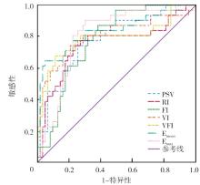
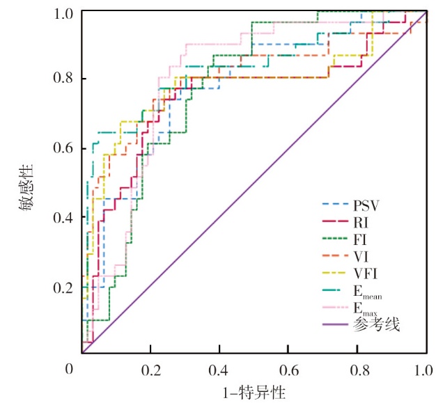
目的 分析多模态超声成像技术[常规二维超声联合经阴道彩色多普勒超声(TVCDS)、三维能量多普勒超声(3D-PDUS)、剪切波弹性成像(SWE)]鉴别诊断宫颈良恶性病变的应用价值。方法 回顾性选取2024年7月至2025年7月于秦皇岛市第一医院就诊的94例宫颈病变患者作为研究对象。评估各超声及多模态超声成像技术对宫颈良恶性病变的诊断效能;比较宫颈炎、宫颈上皮内瘤变、宫颈癌患者TVCDS参数收缩期峰值流速(PSV)、阻力指数(RI),3D-PDUS参数血流指数(FI)、血管指数(VI)、血管-血流指数(VFI),SWE参数平均弹性值(Emean)、最大弹性值(Emax)差异。采用受试者操作特征(ROC)曲线评估各超声参数对宫颈良恶性病变的诊断价值。结果 94例宫颈病变患者中良性病变63例(宫颈炎38例、宫颈上皮内瘤变25例),恶性病变31例。以病理学检查结果为金标准,常规二维超声、TVCDS、3D-PDUS、SWE、多模态超声成像诊断宫颈良恶性病变的Kappa值分别为0.333、0.534、0.669、0.742、0.976,敏感性分别为67.74%(21/31)、74.19%(23/31)、80.65%(25/31)、87.10%(27/31)、100%(31/31),特异性分别为68.25%(43/63)、80.95%(51/63)、87.30%(55/63)、88.89%(56/63)、98.41%(62/63),准确性分别为68.09%(64/94)、78.72%(74/94)、85.11%(80/94)、88.30%(83/94)、98.94%(93/94),阳性预测值分别为51.22%(21/41)、65.71%(23/35)、75.76%(25/33)、79.41%(27/34)、96.88%(31/32),阴性预测值分别为81.13%(43/53)、86.44%(51/59)、90.16%(55/61)、93.33%(56/60)、100%(62/62),3D-PDUS、SWE诊断宫颈良恶性病变的特异性、准确性、阳性预测值均高于常规二维超声检查(均P<0.05),多模态超声成像诊断宫颈良恶性病变的敏感性、特异性、准确性、阳性预测值、阴性预测值均高于常规二维超声、TVCDS、3D-PDUS、SWE单一检查(均P<0.05)。宫颈炎、宫颈上皮内瘤变、宫颈癌患者TVCDS参数PSV分别为(10.35±2.21)、(13.84±2.61)、(17.29±3.07)cm/s,RI分别为0.72±0.20、0.63±0.17、0.52±0.12,差异均有统计学意义(F=59.86,P<0.001;F=11.93,P<0.001);3D-PDUS参数FI分别为44.48(37.79,49.45)、54.05(40.13,65.40)、57.78(45.54,71.03),VI分别为1.20(0.87,1.89)、3.27(1.54,4.24)、5.23(2.98,14.77),VFI分别为0.57(0.25,0.96)、2.04(0.74,2.85)、9.31(5.25,19.82),差异均有统计学意义(U=16.60,P<0.001;U=55.97,P<0.001;U=52.18,P<0.001);SWE参数Emean分别为(20.81±5.61)、(30.05±9.06)、(80.39±9.27)kPa,Emax分别为(27.16±7.14)、(35.53±8.53)、(90.13±8.88)kPa,差异均有统计学意义(F=527.62,P<0.001;F=568.76,P<0.001);其中宫颈上皮内瘤变、宫颈癌患者PSV、FI、VI、VFI、Emean、Emax均高于宫颈炎患者,RI均低于宫颈炎患者,且宫颈癌患者PSV、VI、VFI、Emean、Emax均高于宫颈上皮内瘤变患者,RI低于宫颈上皮内瘤变患者(均P<0.05)。ROC曲线分析显示,PSV、RI、FI、VI、VFI、Emean、Emax诊断宫颈良恶性病变的曲线下面积分别为0.78、0.75、0.78、0.81、0.79、0.83、0.81。结论 多模态超声成像技术可显著提高宫颈良恶性病变的诊断准确性,且参数PSV、RI、FI、VI、VFI、Emean、Emax对宫颈良恶性病变均具有一定的诊断价值。


关键词: 宫颈肿瘤, 宫颈疾病, 超声检查,多普勒, 弹性成像技术

Abstract:

Objective To analyze the application value of multimodal ultrasound imaging [routine two-dimensional ultrasound combined with transvaginal color Doppler sonography (TVCDS),three-dimensional power Doppler ultrasound (3D-PDUS),shear wave elastography (SWE)] in the differential diagnosis of benign and malignant cervical lesions. Methods A total of 94 patients with cervical lesions treated at First Hospital of Qinhuangdao from July 2024 to July 2025 were retrospectively enrolled as research objects. Diagnostic efficiency of different ultrasound and multimodal ultrasound imaging technologies for benign and malignant cervical lesions was evaluated. The differences in TVCDS parameters [peak systolic velocity (PSV),resistance index (RI)],3D-PDUS parameters [flow index (FI),vascularization index (VI),vascularization-flow index (VFI)] and SWE parameters [mean elasticity value (Emean),maximum elasticity value (Emax)] among patients with cervicitis,cervical intraepithelial neoplasia and cervical cancer were compared. The diagnostic value of different ultrasound parameters for benign and malignant cervical lesions was evaluated by receiver operator characteristic (ROC) curves. Results Among the 94 patients with cervical lesions,there were 63 cases with benign lesions (38 cases with cervicitis,25 cases with cervical intraepithelial neoplasia) and 31 cases with malignant lesions. Taking the pathological examination results as the golden standard,Kappa values of two-dimensional ultrasound,TVCDS,3D-PDUS,SWE and multimodal ultrasound imaging in the diagnosis of benign and malignant cervical lesions were 0.333,0.534,0.669,0.742,0.976,sensitivity were 67.74% (21/31),74.19% (23/31),80.65% (25/31),87.10% (27/31),100% (31/31),specificity were 68.25% (43/63),80.95% (51/63),87.30% (55/63),88.89% (56/63),98.41% (62/63),accuracy were 68.09% (64/94),78.72% (74/94),85.11% (80/94),88.30% (83/94),98.94% (93/94),positive predictive values were 51.22% (21/41),65.71% (23/35),75.76% (25/33),79.41% (27/34),96.88% (31/32),negative predictive values were 81.13% (43/53),86.44% (51/59),90.16% (55/61),93.33% (56/60),100% (62/62),respectively. The specificity,accuracy and positive predictive value of 3D-PDUS and SWE in the diagnosis of benign and malignant cervical lesions were higher than those of routine two-dimensional ultrasound (all P<0.05). The sensitivity,specificity,accuracy,positive predictive value and negative predictive value of multimodal ultrasound in the diagnosis of benign and malignant cervical lesions were higher than those of routine two-dimensional ultrasound,TVCDS,3D-PDUS,SWE as single examinations (all P<0.05). The PSV of the TVCDS parameters in patients with cervicitis,cervical intraepithelial neoplasia and cervical cancer were (10.35±2.21),(13.84±2.61),(17.29±3.07) cm/s,RI were 0.72±0.20,0.63±0.17,0.52±0.12,respectively,with statistically significant differences (F=59.86,P<0.001; F=11.93,P<0.001). The FI of the 3D-PDUS parameters were 44.48 (37.79,49.45),54.05 (40.13,65.40),57.78 (45.54,71.03),VI were 1.20 (0.87,1.89),3.27 (1.54,4.24),5.23 (2.98,14.77),VFI were 0.57 (0.25,0.96),2.04 (0.74,2.85),9.31 (5.25,19.82),respectively,with statistically significant differences (U=16.60,P<0.001; U=55.97,P<0.001; U=52.18,P<0.001). The Emean of the SWE parameters were (20.81±5.61),(30.05±9.06),(80.39±9.27) kPa,Emax were (27.16±7.14),(35.53±8.53),(90.13±8.88) kPa,respectively,with statistically significant differences (F=527.62,P<0.001; F=568.76,P<0.001). PSV,FI,VI,VFI,Emean and Emax in patients with cervical intraepithelial neoplasia and cervical cancer were higher than those with cervicitis,while RI was lower than that with cervicitis patients. PSV,VI,VFI,Emean and Emax in patients with cervical cancer were higher than those with cervical intraepithelial neoplasia,while RI was lower than that with cervical intraepithelial neoplasia (all P<0.05). ROC curve analysis showed that,area under the curve values of PSV,RI,FI,VI,VFI,Emean and Emax in the diagnosis of benign and malignant cervical lesions were 0.78,0.75,0.78,0.81,0.79,0.83 and 0.81,respectively. Conclusions Multimodal ultrasound imaging can significantly improve diagnostic accuracy of benign and malignant cervical lesions. PSV,RI,FI,VI,VFI,Emean and Emax all have certain diagnostic value in benign and malignant cervical lesions.

Key words: Uterine cervical neoplasms, Uterine cervical diseases, Ultrasonography,Doppler, Elasticity imaging techniques