国际肿瘤学杂志 ›› 2025, Vol. 52 ›› Issue (1): 23-30.doi: 10.3760/cma.j.cn371439-20240607-00003

• 论著 • 上一篇    下一篇

RRM2通过调控CDK1对胃癌细胞恶性生物学行为及有氧糖酵解的影响

谭荣坚1, 欧雯婷2, 翟嘉伟1, 全祯豪1, 孙利君1, 周才进1()   

  1. 1广东医科大学附属医院胃肠外科,湛江 524000
    2广东医科大学附属医院消化肿瘤内科,湛江 524000
  • 收稿日期:2024-06-07 修回日期:2024-11-24 出版日期:2025-01-08 发布日期:2025-01-21
  • 通讯作者: 周才进 E-mail:zhoucaijin2021@126.com
  • 基金资助:
    湛江市科技发展专项资金竞争性分配项目(2022A01174);湛江市非资助科技攻关计划(2022B01096)

Effects of RRM2 on malignant biological behavior and aerobic glycolysis of gastric cancer cells by regulating CDK1

Tan Rongjian1, Ou Wenting2, Zhai Jiawei1, Quan Zhenhao1, Sun Lijun1, Zhou Caijin1()   

  1. 1Department of Gastrointestinal Surgery, Affiliated Hospital of Guangdong Medical University, Zhanjiang 524000, China
    2Department of Digestive Oncology, Affiliated Hospital of Guangdong Medical University, Zhanjiang 524000, China
  • Received:2024-06-07 Revised:2024-11-24 Online:2025-01-08 Published:2025-01-21
  • Contact: Zhou Caijin E-mail:zhoucaijin2021@126.com
  • Supported by:
    Zhanjiang Science and Technology Development Special Fund Competitive Allocation Project(2022A01174);Zhanjiang Non-Funded Science and Technology Research Program(2022B01096)

摘要:

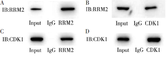
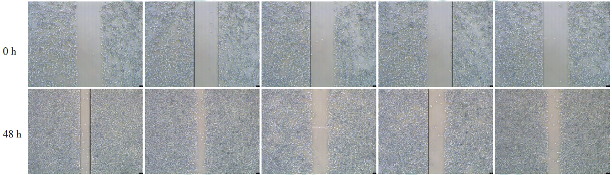
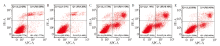
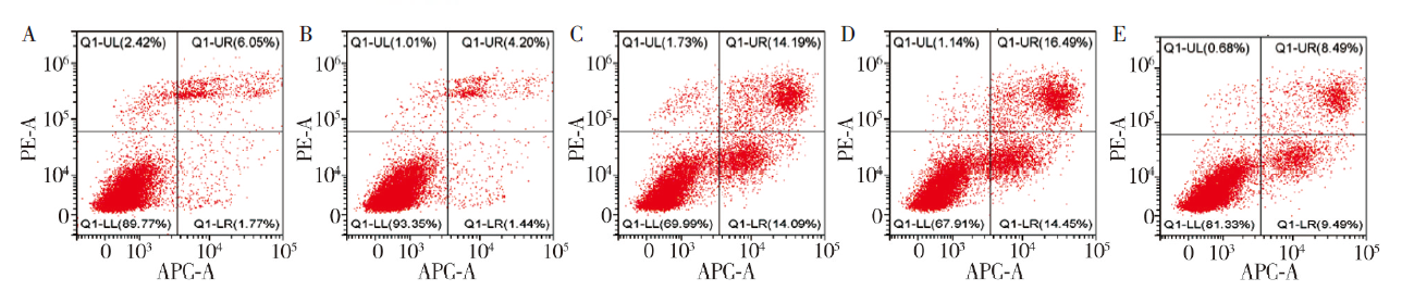
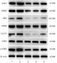
目的 探究核糖核苷酸还原酶调节亚基M2(RRM2)通过调控周期蛋白依赖性激酶(CDK)1对胃癌细胞恶性生物学行为及有氧糖酵解的影响。方法 将人胃癌MKN-45细胞分为si-NC组(转染空白片段)、CoCl2+si-NC组(低氧环境转染空白片段)、CoCl2+si-RRM2组(低氧环境沉默RRM2)、CoCl2+si-RRM2+pcDNA3.1 NC组(低氧环境沉默RRM2+空白载体)、CoCl2+si-RRM2+pcDNA3.1 CDK1组(低氧环境沉默RRM2+过表达CDK1)。实时荧光定量反转录PCR分析RRM2和CDK1 mRNA的相对表达量;免疫共沉淀分析RRM2和CDK1蛋白间的相互作用;MTT法检测细胞增殖活性;细胞划痕实验检测细胞迁移距离;流式细胞术检测细胞凋亡水平;三磷酸腺苷(ATP)和葡萄糖试剂盒检测ATP产生和葡萄糖消耗情况;蛋白质印迹法检测ENO1、RRM2、HK2、PKM2、GLUT1及p-CDK1/CDK1蛋白表达情况。结果 实时荧光定量反转录PCR结果显示,si-NC组、CoCl2+si-NC组和CoCl2+si-RRM2组CDK1 mRNA相对表达量分别为1.01±0.15、1.30±0.06、0.51±0.18,RRM2 mRNA相对表达量分别为1.03±0.32、1.59±0.28、0.44±0.17,差异均有统计学意义(F=25.52,P=0.001;F=14.47,P=0.005);与si-NC组比较,CoCl2+si-NC组细胞RRM2和CDK1 mRNA表达均较高;与si-NC组、CoCl2+si-NC组比较,CoCl2+si-RRM2组细胞RRM2和CDK1 mRNA表达均较低(均P<0.05)。免疫共沉淀结果显示,RRM2与CDK1之间存在相互作用。MTT法、细胞划痕实验和流式细胞术结果显示,si-NC组、CoCl2+si-NC组、CoCl2+si-RRM2组、CoCl2+si-RRM2+pcDNA3.1 NC组和CoCl2+si-RRM2+pcDNA3.1 CDK1组细胞增殖活性分别为1.04±0.01、1.18±0.04、0.84±0.03、0.81±0.03、0.93±0.05,细胞迁移距离分别为(301.83±2.75)、(369.67±0.76)、(176.50±6.38)、(175.83±3.69)、(254.17±1.61)μm,细胞凋亡率分别为8.05%±0.21%、5.75%±0.20%、28.28%±0.04%、30.18%± 1.51%、17.79%±0.22%,差异均有统计学意义(F=73.82,P<0.001;F=1 600.01,P<0.001;F=787.15,P<0.001);与si-NC组和CoCl2+si-NC组比较,CoCl2+si-RRM2组、CoCl2+si-RRM2+pcDNA3.1 NC组和CoCl2+si-RRM2+pcDNA3.1 CDK1组细胞增殖、迁移能力较弱,细胞凋亡率较高(均P<0.05);与CoCl2+si-RRM2+pcDNA3.1 NC组比较,CoCl2+si-RRM2+pcDNA3.1 CDK1组细胞增殖、迁移能力较强,细胞凋亡率较低(均P<0.05)。ATP和葡萄糖检测结果显示,5组细胞ATP产生量和葡萄糖消耗量差异均有统计学意义(F=12.53,P<0.001;F=19.21,P<0.001);与si-NC组比较,CoCl2+si-RRM2+pcDNA3.1 CDK1组细胞葡萄糖消耗量较少(P<0.05);与CoCl2+si-NC组比较,CoCl2+si-RRM2+pcDNA3.1 CDK1组细胞ATP产生量和葡萄糖消耗量均较少(均P<0.05);与CoCl2+si-RRM2+pcDNA3.1 NC组比较,CoCl2+si-RRM2+pcDNA3.1 CDK1组细胞ATP产生量和葡萄糖消耗量均较多(均P<0.05)。蛋白质印迹法检测显示,5组细胞ENO1、RRM2、HK2、PKM2、GLUT1、p-CDK1/CDK1蛋白表达差异均有统计学意义(均P<0.001);与si-NC组和CoCl2+si-NC组比较,CoCl2+si-RRM2+pcDNA3.1 CDK1组细胞ENO1、RRM2、HK2、PKM2、GLUT1和p-CDK1/CDK1蛋白表达均较低(均P<0.05);与CoCl2+si-RRM2+pcDNA3.1 NC组比较,CoCl2+si-RRM2+pcDNA3.1 CDK1组细胞ENO1、RRM2、PKM2、GLUT1和p-CDK1/CDK1蛋白表达均较高(均P<0.05)。结论 沉默RRM2可通过调控CDK1抑制胃癌细胞恶性生物学行为及有氧糖酵解的发生。

关键词: 胃肿瘤, 核苷酸还原酶类, 细胞周期蛋白质依赖性激酶类

Abstract:

Objective To investigate the effect of ribonucleotide reductase regulatory subunit M2 (RRM2) on the malignant biological behavior and aerobic glycolysis of gastric cancer cells by regulating cyclin-dependent kinase (CDK) 1. Methods Human gastric cancer MKN-45 cells were divided into si-NC group (transfected with blank fragment), CoCl2+si-NC group (hypoxia control transfected with blank fragment), CoCl2+si-RRM2 group (hypoxia with RRM2 silencing), CoCl2+si-RRM2+pcDNA3.1 NC group (hypoxia with RRM2 silencing and blank vector) and CoCl2+si-RRM2+pcDNA3.1 CDK1 group (hypoxia with RRM2 silencing and CDK1 overexpression). The mRNA relative expression levels of RRM2 and CDK1 were analyzed by real time fluorescent quantitative reverse transcription PCR. Co-immunoprecipitation (CoIP) was used to analyze the interaction between RRM2 and CDK1 protein. MTT assay was used to analyze the proliferation activity of cells. The cell migration distance was detected by cell scratch assay. Cell apoptosis was detected by flow cytometry. Adenosine triphosphate (ATP) and glucose kit were used to detect ATP production and glucose consumption. The protein expressions of ENO1, RRM2, HK2, PKM2, GLUT1 and p-CDK1/CDK1 were detected by Western blotting. Results Real time fluorescent quantitative reverse transcription PCR results showed that the relative expression levels of CDK1 mRNA in si-NC group, CoCl2+si-NC group and CoCl2+si-RRM2 group were 1.01±0.15, 1.30±0.06 and 0.51±0.18, and the relative expression levels of RRM2 mRNA were 1.03±0.32, 1.59±0.28 and 0.44±0.17, respectively, and there were statistically significant differences (F=25.52, P=0.001; F=14.47, P=0.005). The mRNA expressions of RRM2 and CDK1 in CoCl2+si-NC group were higher than those in si-NC group. Compared with the si-NC group and the CoCl2+si-NC group, the mRNA expressions of RRM2 and CDK1 were lower in the CoCl2+si-RRM2 group (all P<0.05). CoIP results showed that there was interaction between RRM2 and CDK1. MTT assay, cell scratch assay and flow cytometry showed that the cell proliferation activity of si-NC group, CoCl2+si-NC group, CoCl2+si-RRM2 group, CoCl2+si-RRM2+pcDNA3.1 NC group and CoCl2+si-RRM2+pcDNA3.1 CDK1 group were 1.04±0.01, 1.18±0.04, 0.84±0.03, 0.81±0.03 and 0.93±0.05, respectively. The cell migration distances were (301.83±2.75), (369.67±0.76), (176.50±6.38), (175.83±3.69), (254.17±1.61) μm, respectively. The apoptosis rates were 8.05%±0.21%, 5.75%± 0.20%, 28.28%±0.04%, 30.18%±1.51% and 17.79%±0.22%, respectively, all with statistically significant differences (F=73.82, P<0.001; F=1 600.01, P<0.001; F=787.15, P<0.001). Compared with the si-NC group and CoCl2+si-NC group, the proliferation and migration ability of cells in the CoCl2+si-RRM2 group, CoCl2+si-RRM2+pcDNA3.1 NC group and CoCl2+si-RRM2+pcDNA3.1 CDK1 group were weaker, and the apoptosis rates were higher (all P<0.05). Compared with the CoCl2+si-RRM2+pcDNA3.1 NC group, the proliferation and migration ability of cells in the CoCl2+si-RRM2+pcDNA3.1 CDK1 group were stronger, and the apoptosis rate was lower (all P<0.05). The results of ATP and glucose detection showed that there were statistically significant differences in the amount of ATP production and glucose consumption among the above five groups (F=12.53, P<0.001; F=19.21, P<0.001). Compared with the si-NC group, the glucose consumption of cells was lower in the CoCl2+si-RRM2+pcDNA3.1 CDK1 group (P<0.05). Compared with the CoCl2+si-NC group, the ATP production and glucose consumption of cells in the CoCl2+si-RRM2+pcDNA3.1 CDK1 group were lower (both P<0.05). Compared with the CoCl2+si-RRM2+pcDNA3.1 NC group, the ATP production and glucose consumption of the CoCl2+si-RRM2+pcDNA3.1 CDK1 group were higher (both P<0.05). Western blotting showed that there were statistically significant differences in the protein expressions of ENO1, RRM2, HK2, PKM2, GLUT1, and p-CDK1/CDK1 among the above five groups (all P<0.001). Compared with the si-NC group and the CoCl2+si-NC group, the protein expressions of ENO1, RRM2, HK2, PKM2, GLUT1 and p-CDK1/CDK1 in the CoCl2+si-RRM2+pcDNA3.1 CDK1 group were lower (all P<0.05). Compared with the CoCl2+si-RRM2+pcDNA3.1 NC group, the protein expressions of ENO1, RRM2, PKM2, GLUT1 and p-CDK1/CDK1 in the CoCl2+si-RRM2+pcDNA3.1 CDK1 group were higher (all P<0.05). Conclusions Silencing RRM2 can inhibit the malignant biological behavior of gastric cancer cells and the occurrence of aerobic glycolysis by regulating CDK1.

Key words: Stomach neoplasms, Ribonucleotide reductases, Cyclin-dependent kinases